Home platform cell model architectures
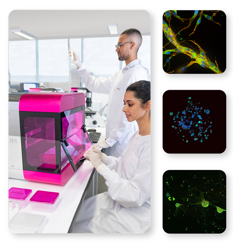
Scientists in a laboratory working with the RASTRUM™ platform, loading a well plate for 3D cell model generation. Adjacent fluorescence microscopy images showcase diverse 3D cell model architectures optimized for applications such as imaging, screening, and cell extraction.

Ready-to-use cell model architectures optimized for your applications

Empower your research with a range of 3D cell model architectures tailored to your needs. Seamlessly integrate these flexible architectures into various well-plate formats and applications, including imaging, screening, and cell extraction.
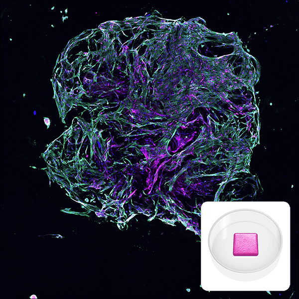
Fluorescent microscopy image of a lung fibroblast model printed with RASTRUM, showing collagen (magenta), actin (green), and cell nuclei (blue). Inset displays a schematic of the Imaging Model architecture, optimized for high-content imaging and minimal cell usage.

Imaging Model

A small single-matrix architecture optimized for fast imaging and minimal cell usage. This model ensures clear imaging and compatibility with standard techniques, enabling seamless integration into your workflows. Compatible with 96-well plates on RASTRUM™ and RASTRUM™ Allegro.

Applications

  • Brightfield and immunofluorescence imaging
  • Drug screening
  • Biochemical assays

Features

  • Thin architecture for rapid, high-content imaging
  • Compact design minimizes the number of cells required while maintaining reproducibility


Image depicts a lung fibroblast model made with RASTRUM. Primary normal human lung fibroblasts (NHLFs) were printed using RASTRUM’s Imaging Model architecture. Cells were cultured for 7 days and immunostaining was performed to visualize collagen I (magenta), alpha smooth muscle actin (green) and cell nuclei (blue). Inset: Imaging Model architecture illustration. 

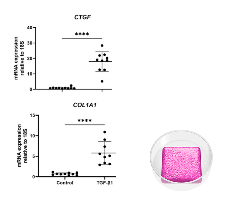
Graph showing increased gene expression of COL1A1 and CTGF in a liver co-culture model created with RASTRUM after TGF-β1 treatment. Inset displays a schematic of the Large Plug Model, optimized for bulk cell analysis and high data yield in DNA, RNA, and protein studies.

Large Plug Model

A single-matrix architecture tailored for expansion and bulk downstream analyses, delivering the cellular material you need for comprehensive studies and high-throughput applications. Compatible with 96-well plates on RASTRUM and RASTRUM Allegro.

Applications

  • DNA, RNA, and protein analysis
  • Flow cytometry
  • Omics analyses

Features

  • Larger matrix architecture for increased cellular material
  • Optimized for workflows requiring bulk analysis and high data yield


Image depicts analysis of a liver co-culture created with RASTRUM. Immortalized hepatocyte and stellate cells were printed in a Large Plug Model and cultured for 5 days before treatment with TGF-β1. On Day 7 cells were retrieved from the matrix and RNA isolated. Gene expression of profibriotic markers for Collagen-type 1 (COL1A1) and connective tissue growth factor (CTGF) were shown to be significantly increased following TGF-ꞵ1 treatment. Inset: Large Plug Model architecture illustration. 

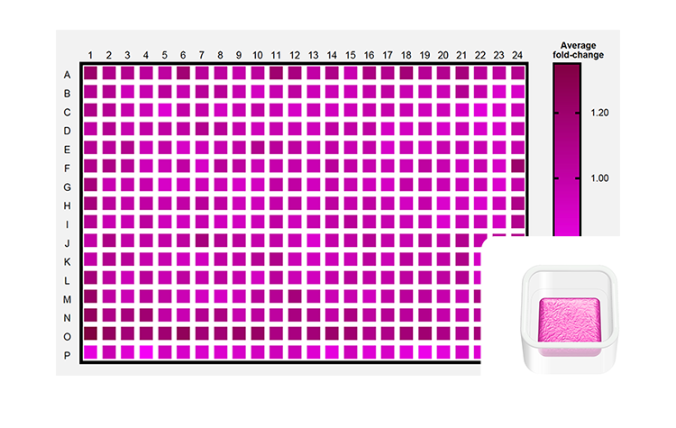
Heatmap showing drug sensitivity assay results for MCF-7 cells printed in a Screening Model architecture across 384-well plates. Inset displays a schematic of the Screening Model, designed for high-throughput workflows, drug screening, and cytotoxicity assays.

Screening Model

A single-matrix architecture optimized for high-throughput workflows, enabling efficient data collection and scalable experimentation. Compatible with 384-well plates on RASTRUM Allegro.

Applications

  • Drug screening
  • Cytotoxicity assays
  • Mechanistic studies


Features

  • Optimized for assay flexibility and adaptability
  • Consistent and reproducible results across screening workflows


Image depicts consistent MCF-7 drug sensitivities at scale. MCF-7 cells printed in the Screening Model architecture in wells of twelve 384-well plates across two independent PrintRuns. Low post-print and drug treatment CVs were observed. Inset: Screening Model architecture illustration. 

Fluorescent microscopy image of a 3D metastatic mammary adenocarcinoma culture printed with RASTRUM in a High-Throughput Model. Stained nuclei (blue), endoplasmic reticulum (green), and actin (yellow) highlight cell morphology. Inset shows a schematic of the High-Throughput Model for scalable assays.

High-Throughput Model

A single-matrix architecture optimized for high-throughput workflows in 384-well plates, enabling efficient data collection and scalable experimentation. Compatible with 384-well plates on RASTRUM. 

Applications

  • Drug screening
  • High-content imaging
  • Biochemical assays

Features

  • Consistent and reproducible 3D models in 384-well plate format
  • Streamlined for large-scale assays and automated workflows


Image depicts a 3D culture of a metastatic mammary adenocarcinoma. Matrix cultures with MDA-MB-231 cells were generated with RASTRUM using the High-Throughput Model architecture, and grown for 7 days in 384 well plates. Morphology staining was applied for high content phenotypic imaging using the PhenoVue Cell Painting Kit: nuclei (blue, Hoechst), endoplasmic reticulum (green, concanavalin A), and actin (yellow , phalloidin). Inset: High-Throughput Model architecture illustration.

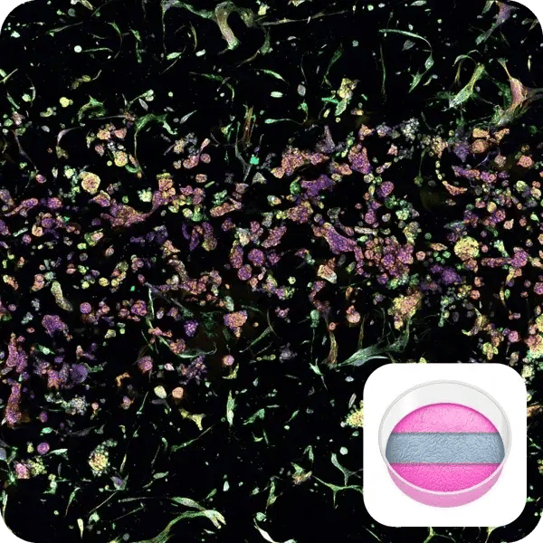
Fluorescent microscopy image of tumor-fibroblast cell interactions in a RASTRUM Triple Matrix Model. Cells stained for nuclei (blue), endoplasmic reticulum (green), and actin (yellow) show interactions across three distinct matrix compartments. Inset displays a schematic of the Triple Matrix Model.

Triple Matrix Model

A triple-matrix design for recapitulating tissue architecture and studying complex cell interactions. Compatible with 96-well plates on RASTRUM and RASTRUM Allegro. 

Applications

  • Migration and invasion
  • Tissue architecture recapitulation
  • Cell signaling and interaction
  • Wound healing and gap closure assays

Features

  • Co-culture three distinct cell types in side-by-side compartments
  • Independently define matrix stiffness, composition, and cellular components
  • Visualize cell movement and interaction between compartments
  • Simulate gap closure for wound healing and migration studies


Image illustrates tumor-fibroblast cell interactions within RASTRUM’s Triple Matrix Model architecture. Normal human lung fibroblasts (NHLFs) printed in the top and bottom matrix compartments of the model architecture interact with A549 lung adenocarcinoma cells growing within the middle compartment. Cells were labelled using the PhenoVue Cell Painting Kit: nuclei (blue, Hoechst), endoplasmic reticulum (green, concanavalin A), and actin (yellow , phalloidin), at day 7 post-printing. Inset: Triple Matrix Model architecture illustration. 

Fluorescent microscopy image of tumor microenvironment modeling using RASTRUM Dual Matrix Model. Lung cancer cells (left) and fibroblasts (right) are separated by defined matrix boundaries, with nuclei (blue), endoplasmic reticulum (green), and actin (yellow) staining. Inset shows a schematic of the Dual Matrix Model.

Dual Matrix Model

A dual-matrix architecture optimized for recapitulating tissue architecture and studying cellular interactions within a 3D environment. Compatible with 96-well plates on RASTRUM and RASTRUM Allegro. 

Applications

  • Migration and invasion studies
  • Tissue architecture recapitulation
  • Cell signaling and interaction

Features

  • Enables imaging of cell signaling and interaction with clarity
  • Provides 3D models with defined and reproducible tissue architecture
  • Facilitates advanced studies on cellular behavior in complex environments


Image illustrates tumor microenvironment with spatial separation in RASTRUM’s Dual Matrix Model architecture. Lung cancer (A549) cells (left) and normal human lung fibroblast (right) were printed in a Dual Matrix Model architecture on RASTRUM and imaged after seven days in vitro. Dotted lines indicate the boundaries between the matrices. Cells were labelled using the PhenoVue Cell Painting Kit: nuclei (blue, Hoechst), endoplasmic reticulum (green, concanavalin A), and actin (yellow , phalloidin). Inset: Dual Matrix Model architecture illustration. 

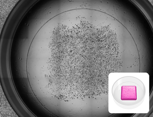
Black and white microscopy image of an MCF-7 cancer cell culture printed in a RASTRUM Removable Model within a 24-well plate, designed for implantation and downstream analyses. The coverslip enables easy removal of the intact matrix. Inset shows a schematic of the Removable Model architecture.

Removable Model

A large removable architecture designed for 24-well plates and printed on coverslips, offering exceptional versatility for implantation and downstream analyses. Compatible with 24-well plates on RASTRUM and RASTRUM Allegro. 

Applications

  • Implantation studies
  • Immunohistochemistry
  • Spatial biology

Features

  • Easy removal for microscopic imaging
  • Simplified handling for embedding and sectioning
  • Ideal for workflows requiring intact tissue preparation


Image shows cancer cell culture in a 24-well plate format in preparation for immunohistochemistry. MCF-7 cells were printed in the RASTRUM Removable Model architecture and imaged after 3 days. The coverslip facilitates easy removal of the intact matrix for downstream applications. Inset: Removal Model architecture illustration.

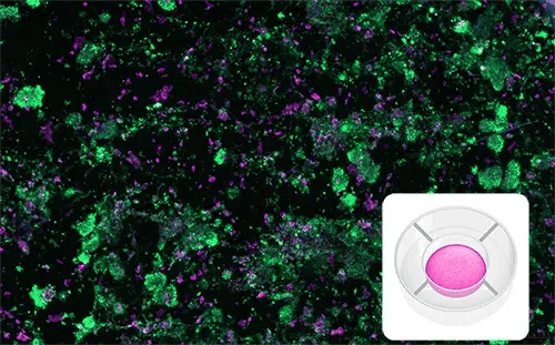
Fluorescent microscopy image of an immuno-oncology model using RASTRUM Mematix Model, showing CD45+ immune cells (green) interacting with Pan-CK+ cancer cells (magenta) after six days. Designed for migration and signaling studies. Inset displays a schematic of the Mematix Model architecture.

Mematix Model

A single-matrix architecture for transwell insert membranes, enabling advanced migration and signaling studies. Compatible with 96-well plates on RASTRUM and RASTRUM Allegro. 

Applications

  • Evaluation of cell movement and migration
  • Paracrine signaling studies

Features

  • Supports two separate media chambers for controlled chemokine or chemoattractant gradients
  • Allows migration of cells through the membrane for advanced migration studies


Immuno-oncology model generated with RASTRUM, showcasing interactions between PBMCs and A549 cells in a Mematix Model architecture, imaged after 6 days. CD45+ immune cells (green) are observed interacting with Pan-CK+ cancer cells (magenta). Inset: Mematix Model architecture illustration. 

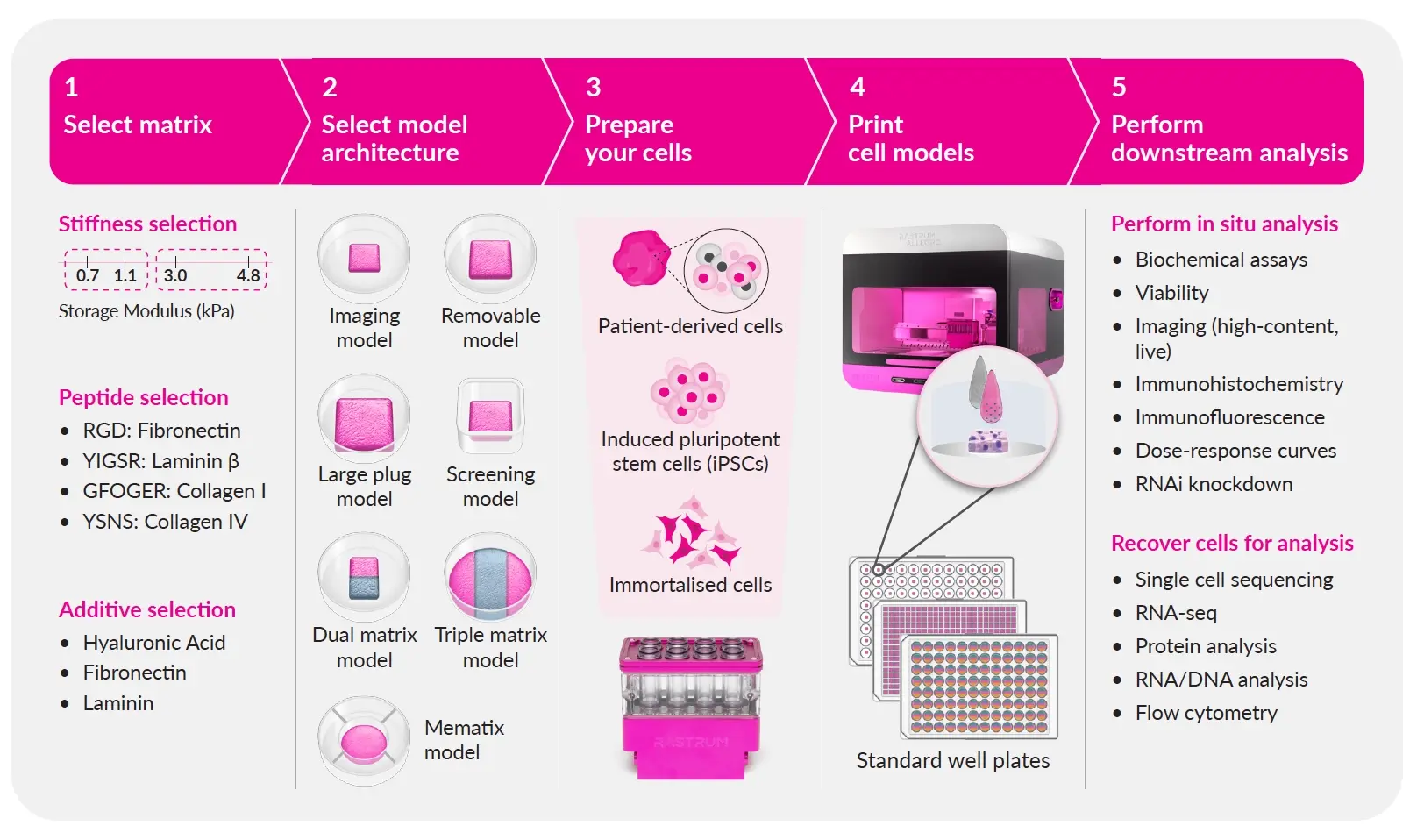
Step-by-step process for generating 3D cell models with RASTRUM Allegro: select matrix, choose architecture, prepare cells, print models, and perform downstream analysis for high-throughput drug discovery and biomedical research.

RASTRUM technology is designed to streamline your 3D cell model development from start to finish, delivering biologically relevant results with efficiency and precision. Simply point, click, and experiment with our cloud-based guided experimental design, ready-to-use protocols, and expert assistance.   

  1. Select your matrix: Choose from our large library of flexible, tunable, xeno-free matrices to mimic the tissue microenvironment to suit your research.
  2. Select your cell model architecture: Choose the structure and configuration of your cell model from our options to suit your experimental needs.
  3. Prepare your cells: Follow a simple protocol to prepare your patient-derived, iPSC or immortalized cells
  4. Print your cell models: Utilize RASTRUM's drop-on-demand bioprinting technology to rapidly and reproducibly generate your 3D cell models.
  5. Perform your downstream analysis: Grow your cells and easily integrate cell model analysis with existing downstream workflows and readouts.
FAQs
What is an inert base layer in a RASTRUM 3D model?
An inert base layer is a non-functionalized PEG-based gel layer printed between the tissue culture plastic and the 3D cell model. It prevents cells from attaching to the bottom of the well and, when printed correctly, prevents 2D growth on the well surface.
Why does RASTRUM use an inert base layer?
RASTRUM uses an inert base layer in many architectures to prevent cells from attaching to the bottom of the well. By separating the 3D cell model from the plastic surface, the inert base layer helps avoid 2D monolayer formation and supports a true 3D culture environment.
What are RASTRUM cell model architectures?
RASTRUM cell model architectures are predefined 3D model formats that control how cell-loaded matrices are arranged within a well. Different architectures support different experimental goals, such as imaging, screening, co-culture, migration, invasion, omics, histology, or high-throughput workflows.
How do researchers choose the right RASTRUM architecture for an experiment?
Researchers should choose a RASTRUM architecture based on the biological question, plate format, cell number, assay, imaging needs, co-culture design, and downstream analysis. For example, imaging-focused studies may use Imaging Models, while omics or flow cytometry may require Large Plug-style formats.
What is the RASTRUM Imaging Model used for?
The RASTRUM Imaging Model is a small single-matrix architecture designed for fast imaging, minimal cell usage, and compatibility with standard analysis techniques. It can support brightfield imaging, immunofluorescence imaging, high-content screening, biochemical assays, and selected drug-screening workflows where compatible.
What is the RASTRUM Large Plug Model used for?
The RASTRUM Large Plug Model is a single-matrix architecture intended to generate larger amounts of cellular material for expansion and bulk downstream analyses, including DNA, RNA, and protein analysis where compatible. It can also support therapeutic testing, automated imaging, organoid or tumoroid culture, and direct co-culture workflows.
What is the RASTRUM Screening Model used for?
The RASTRUM Screening Model is a single-matrix architecture optimized for scalable 384-well workflows on RASTRUM Allegro. It is intended for applications such as drug screening, cytotoxicity assays, and mechanistic studies where efficient data collection and plate-based scalability are important.
What is the RASTRUM High Throughput Model used for?
The RASTRUM High Throughput Model is a single-matrix architecture optimized for 384-well high-throughput workflows. It supports scalable data collection for applications such as drug screening, high-content screening, high-content imaging, and biochemical assays where the model and assay are compatible.
Which RASTRUM architecture is used for 384-well drug screening?
For RASTRUM Allegro, the Screening Model is the primary 384-well architecture for scalable drug-screening workflows. It is designed for efficient data collection in applications such as dose-response studies, cytotoxicity assays, mechanistic studies, high-content screening, fluorescence staining, imaging, and compatible colorimetric assays.
What is the RASTRUM Dual-Matrix architecture used for?
The RASTRUM Dual-Matrix architecture allows two matrices with similar or different cell and/or matrix composition to be printed in a defined spatial arrangement. It can support migration, invasion, tissue organization, cell signaling, and studies where researchers need to evaluate interactions between adjacent matrix regions.
What is the RASTRUM Triple Matrix - Imaging architecture used for?
The RASTRUM Triple Matrix - Imaging architecture arranges three juxtaposed matrices with different cell and/or matrix compositions to study short-distance interactions in 3D. It can support cell-cell communication, migration, invasion, immuno-oncology, metastasis, neurite outgrowth, automated image analysis, and downstream molecular biology applications.
What is the RASTRUM Mematix Model?
The RASTRUM Mematix Model is a single-matrix architecture for transwell insert membrane workflows. It is intended for advanced migration and signaling studies, including evaluation of cell movement, migration, invasion, chemotaxis, and paracrine signaling depending on the experimental design.
What is the RASTRUM Removable Model used for?
The RASTRUM Removable Model is a large removable architecture designed for 24-well plates and printed on coverslips. It supports workflows where the hydrogel structure needs to be removed for downstream analysis, including histology, spatial biology, high-resolution microscopy, sectioning, and fluorescence or brightfield imaging.
Which RASTRUM architecture is best for RNA, protein, omics, or flow cytometry?
RASTRUM Large Plug architectures are generally better suited to bulk downstream analyses because they can provide more cellular material. Depending on the workflow, they can support DNA, RNA, and protein analysis, flow cytometry, omics workflows, fluorescence staining, and high-magnification imaging.
Which RASTRUM architectures are used for migration, invasion, or spatial co-culture studies?
RASTRUM Dual-Matrix, Triple Matrix, and Mematix architectures are commonly positioned for studies requiring spatial control, migration, invasion, cell-cell communication, paracrine signaling, or tissue-like interfaces. The best architecture depends on whether the experiment requires adjacent compartments, layered interfaces, or transwell-style signaling.