3D cancer models for tumor microenvironment research

Build reproducible 3D cancer models that help researchers study tumor, stromal, immune, matrix, and architecture effects in scalable oncology workflows.

Cancer researchers often know the biology they need to study. The harder problem is building a tumor microenvironment model that is complex enough to matter, reproducible enough to trust, and scalable enough to use in real workflows.

RASTRUMTM helps teams create 3D cell models for cancer research, from exploratory disease biology through drug screening, immuno-oncology, precision medicine, and invasion studies.
Cancer models need to capture more than tumor cell growth

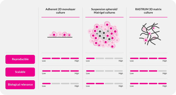
RASTRUM enables scalable, reproducible, and biologically relevant 3D cell models compared to 2D cell culture or suspension cultures.

Cancer is heterogeneous, adaptive, and shaped by its surroundings. Yet many cancer research cell models still simplify the biology that drives progression, treatment resistance, immune evasion, and metastasis.

2D cultures are useful for early experiments, but they often miss important cell-cell and cell-matrix interactions. Animal and in vivo tumor models can provide valuable biology, but they are time-consuming, costly, and difficult to scale for routine screening. In addition, they do not replicate the human immune response well. Manual 3D cancer models can add complexity, but they can also introduce variability that makes results harder to compare and they are generally time consuming. They typically also require a fair amount of material and cells, due to manual handling limits.

Researchers need next generation cancer models that connect biological complexity with workflow control.

RASTRUM supports 3D cancer cell model generation in standard plate formats, helping teams build models around:

  • Tumor-stroma interaction
  • Matrix composition and stiffness
  • Immune-cell infiltration and activation
  • Drug response and treatment resistance mechanisms
  • Patient-derived tumoroids and tumor heterogeneity
  • Migration, invasion, and metastatic behavior
  • Reproducible readouts across plates, runs, and teams
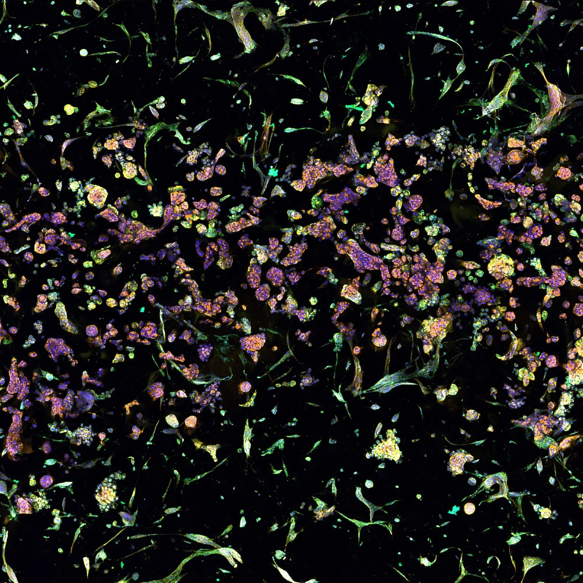
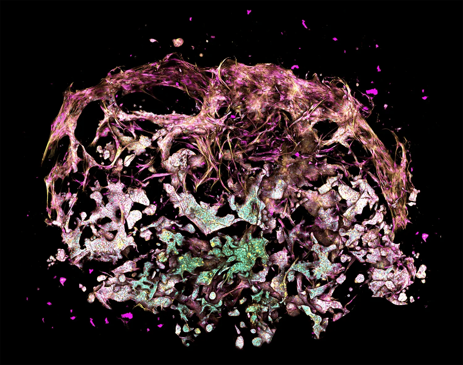
The figure depicts a lung cancer tri-culture model made with RASTRUM Allegro. Primary normal human lung fibroblasts (NHLFs) were printed with primary Human Umbilical Vein Endothelial Cells (HUVECs) and A549 lung adenocarcinoma cells using RASTRUM's Imaging Model architecture. Cells were cultured for 7 days and stained using a PhenoVue Cell Painting Kit (Revvity). Cell nuclei (cyan) were labeled with Hoescht, filamentous actin (yellow) with phalloidin, and membrane glycoproteins (magenta) with concanavalin A.

What RASTRUM enables for 3D cancer research

RASTRUM is designed to help researchers create reproducible, matrix-embedded 3D tumor models with controlled architecture, tunable matrix conditions, and compatibility with downstream cancer biology assays.

Controlled model architecture

Build 3D tumor models in defined formats for imaging, screening, co-culture, cell recovery, spatial organization, migration, invasion, and other cancer research applications.

Tunable matrix environments

Design 3D models around the matrix properties that matter to your question, including stiffness, adhesion cues, remodeling, and extracellular matrix components.

Advanced co-culture capability

Create integrated or spatially defined co-culture models that incorporate tumor cells, fibroblasts, cancer-associated fibroblasts, endothelial cells, immune cells, or other relevant cell types where appropriate.

Scalable workflows for discovery

Move from exploratory 3D cancer biology into repeatable workflows for drug discovery, mechanism studies, immuno-oncology assays, and translational research.

Multiple paths to start

Use Discovery Mode for flexible model development, Validated Solutions for predefined workflows, or Discovery Services when your team needs support developing, running, or transferring a custom 3D disease model.
 

Start with the cancer question you need to answer

Different teams enter the 3D cancer model journey at different points. Some are still evaluating why 3D models matter. Others already know they need a specific tumor microenvironment, immuno-oncology, drug response, patient-derived, or invasion workflow.Use the sections below to find the model direction that fits your research question.

I need to model the tumor microenvironment

 Use 3D tumor microenvironment models to study tumor-stroma crosstalk, matrix effects, treatment resistance, and multicellular interactions. 

I need immuno-oncology 3D models

 Use tumor-immune co-culture models to study immune-cell behavior, infiltration, activation, suppression, and immune-mediated response. 

I need to screen cancer therapeutics in 3D

 Use reproducible 3D tumor screening models to compare response across candidates, combinations, and disease-relevant contexts. 

I need patient-derived or precision medicine research models

 Use patient-derived cells or tumoroids to study heterogeneity, biomarkers, and treatment response in controlled 3D environments. 

I need a migration or invasion model

 Use spatially defined architectures to study cancer cell movement, tumor-stroma interaction, and invasion through controlled 3D environments. 
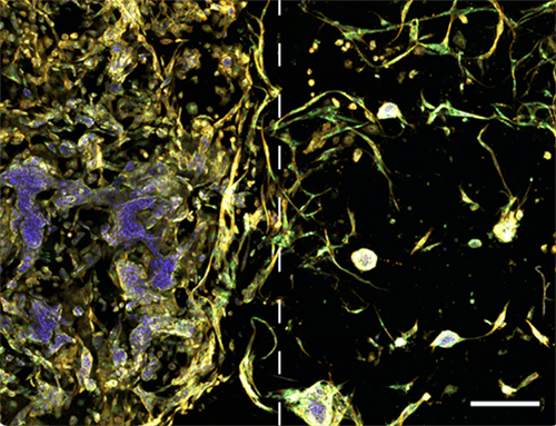
Image illustrates tumor microenvironment with spatial separation in RASTRUM's Dual Matrix Model architecture. Lung cancer (A549) cells (left) and normal human lung fibroblast (right) were printed in a Dual Matrix Model architecture on RASTRUM and imaged after seven days in vitro. Dotted lines indicate the boundaries between the matrices. Cells were labelled using the PhenoVue Cell Painting Kit: nuclei (blue, Hoechst), endoplasmic reticulum (green, concanavalin A), and actin (yellow, phalloidin).

Tumor microenvironment modeling with 3D cancer models

Creating biologically relevant tumor microenvironment models is essential for studying cancer progression, drug resistance, stromal signaling, immune-cell behavior, and therapeutic response. RASTRUM enables researchers to build 3D cancer models that incorporate tumor cells, stromal cells, matrix conditions, and defined architecture into reproducible workflows.

Use this approach when you need to:

  • Recreate tumor-stroma interactions using cancer cells, fibroblasts, CAFs, immune cells, endothelial cells, or matrix components where appropriate
  • Study how matrix stiffness, matrix composition, and hydrogel customization affect tumor phenotype and response
  • Evaluate how stromal components, immune cells, matrix conditions, and treatment resistance shape tumor model behavior
  • Compare how tumor microenvironments influence response across different disease contexts


RASTRUM Matrices can be tuned around the physical and biochemical conditions that shape tumor-stroma biology, including stiffness, adhesion, ECM components, and remodeling.

For teams working in pancreatic cancer, the PDAC Validated Solution provides a predefined 3D tumor-CAF co-culture workflow for studying tumor-stroma interactions, CAF-mediated chemoprotection, and therapeutic response. The Fibroblast Activation Validated Solution provides a defined 3D workflow for studying fibroblast activation, collagen I expression, and stromal biology and testing stroma targeting drugs, that either target stromal signaling pathways or antibody drug conjugates (ADCs) that target stromal cells.
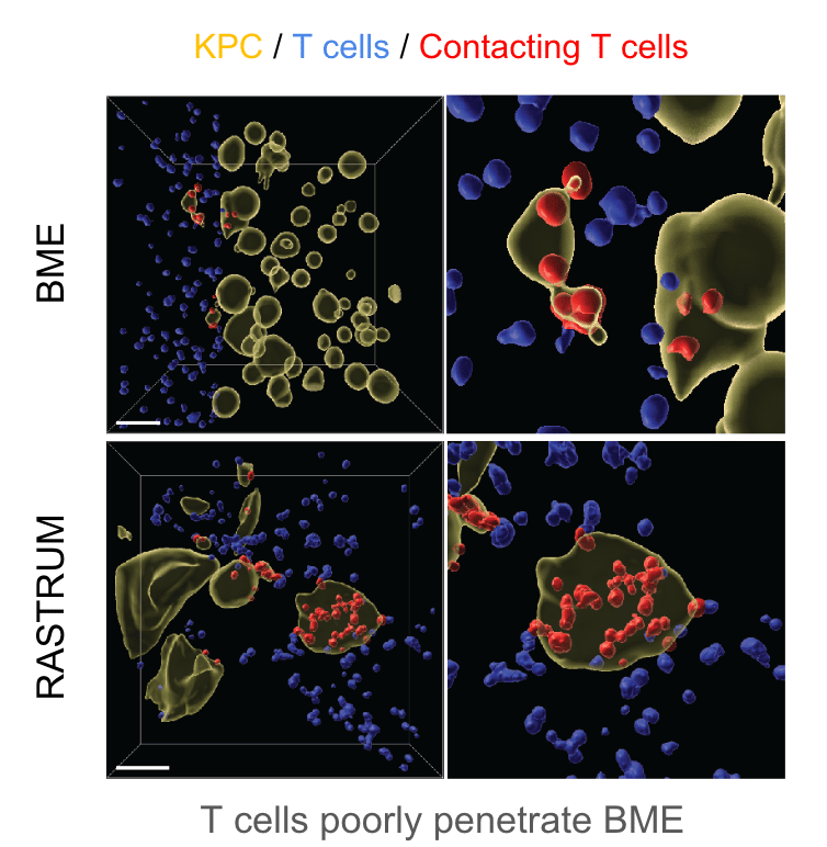
 

The figure depicts pancreatic cancer cells cultured in 4.8kPa matrices for 3 days before 12,000 activated cytotoxic T lymphocytes (CTLs) were added to the media in each well. Imaging and Imaris analysis shows CTL infiltration 2 days post-addition and allows quantification of interactions (red) between cancer cells (yellow) and T cells (blue) in RASTRUM matrices and basement membrane extract (BME).

Immuno-oncology 3D models for tumor-immune interaction studies

Immuno-oncology teams need in vitro immuno-oncology assays that capture more than endpoint tumor killing. In 3D models, immune-cell access, matrix conditions, stromal barriers, cytokine response, activation state, and tumor architecture can all influence interpretation.

RASTRUM supports development of immuno-oncology 3D models and 3D tumor microenvironment co-culture for immuno-oncology research, helping teams study how tumor, stromal, immune, and matrix components shape therapeutic response.

Use this approach when you need to:

  • Model tumor-immune interactions in a defined 3D tumor model
  • Study immune-cell infiltration, activation, suppression, and tumor contact
  • Evaluate CAR-T, checkpoint inhibitor, immune-modulating, or biotherapeutic research questions in biologically relevant 3D models
  • Investigate immune evasion, cytokine signaling, stromal barriers, and resistance mechanisms
  • Compare immune response across tumor microenvironment conditions


For pancreatic cancer and other solid tumor contexts, RASTRUM can support model development where immune-cell infiltration, tumor-immune interaction, and matrix effects are central to the study question. For teams evaluating CAR-T screening in 3D tumor spheroids or related 3D tumor models, the appropriate starting point depends on the cell source, model format, matrix, activation strategy, and readouts.


The figure shows MCF-7 breast cancer cells cultured in 1.1kPa matrices for 6 days before addition of the alkylating agent to wells at 1uM (left) and 100uM (right). Live/dead staining and imaging was performed 24 hours post-addition of the alkylating agent, with live cells shown in green (Calcein-AM) and dead cells in red (EthD-III).

Drug discovery and screening using 3D tumor models

Drug discovery teams need cancer models that can reveal whether therapeutic response holds in more complex biology. A compound that performs well in a simplified system may behave differently when tumor-stroma signaling, matrix effects, immune interactions, or treatment-resistant contexts are introduced.

RASTRUM helps teams generate reproducible 3D tumor models for screening, lead validation, mechanism studies, and cancer therapeutics research.

Use this approach when you need to:

  • Generate standardized 3D tumor models for drug response studies
  • Compare candidate response across defined cancer 3D model conditions
  • Study how tumor microenvironment biology contributes to resistance
  • Evaluate pathway-targeted agents, biologics, ADCs, combinations, or immune-modulating therapies where appropriate
  • Improve confidence in early discovery decisions by reducing variability in 3D model setup
  • Connect cancer research using 3D cell models to downstream imaging, viability, molecular, or functional readouts


RASTRUM can support 3D models for cancer therapeutics across assay development, screening, and lead validation workflows.
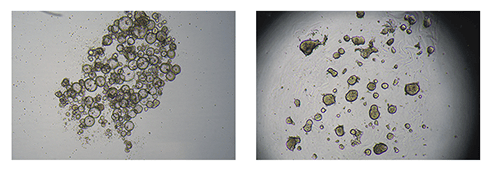
 

The figure depicts bright-field images of patient-derived colorectal cancer cells grown as tumoroids in suspension cultures at day 6 (left) or in Px02 RASTRUM matrices at day 7 (right).

Precision medicine research using patient-derived 3D tumor models

Precision medicine research depends on understanding how tumor biology varies across patients, models, and therapeutic contexts. Patient-derived tumor cells and patient-derived tumoroids can help researchers study heterogeneity, biomarkers, and functional response, but sample availability and workflow variability can limit routine use.

RASTRUM supports reproducible 3D models for patient-relevant cancer studies, including workflows designed around patient-derived cells, tumoroids, and other translational research model systems even with very limited sample quantities, which is often the case with patient samples..

Use this approach when you need to:

  • Build patient-relevant 3D cancer models in controlled matrix environments
  • Study tumor-specific response across cell sources or model conditions
  • Investigate biomarkers linked to sensitivity, resistance, or phenotype
  • Compare patient-derived tumoroids or cells across treatment conditions
  • Support translational research workflows where reproducibility and sample use matter
  • Generate data that helps teams evaluate whether a model is ready for broader assay development


For teams that need a custom patient-derived tumor model or tumor model services, Inventia Discovery Services can help assess feasibility, develop model conditions, run studies, and support transfer when appropriate.
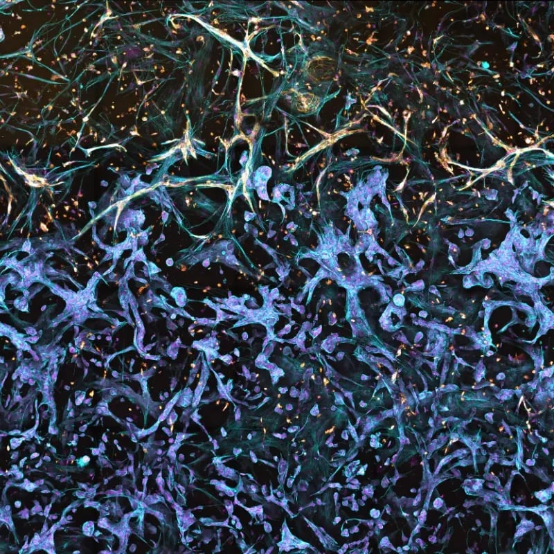
 

The figure depicts a model of the tumor-stroma interface in a Triple Matrix Model made with RASTRUM. Fibroblasts (primary human lung fibroblasts) and endothelial cells (primary HUVECs) were grown next to lung adenocarcinoma cancer cells (A549) and fibroblasts (NHLF) were cultured for 10 days. Endothelial cells were stained with CD31 (yellow) and fibroblasts and endothelial cells were stained with Phalloidin (blue).

3D models for cancer cell migration and invasion

Metastasis and invasion are driven by cell movement, tumor-stroma interaction, matrix remodeling, and microenvironmental cues. Traditional formats often struggle to capture how cancer cells move through or across controlled 3D environments.

RASTRUM supports 3D cancer cell invasion and migration assay development using defined architectures that allow researchers to study cancer cell movement, stromal interaction, and invasion-related biology in reproducible model systems.

Use this approach when you need to:

  • Model cancer cell invasion and metastatic spread in a controlled 3D environment
  • Study epithelial-mesenchymal transition, tumor cell plasticity, migration, and drug resistance
  • Examine how fibroblasts, endothelial cells, immune cells, or matrix composition influence invasion
  • Build tumor-stroma interface models using spatially defined architectures
  • Evaluate potential anti-invasive or anti-metastatic strategies in repeatable 3D workflows


Some teams need a predefined workflow. Others need a custom 3D cancer model built around a specific disease biology, cell source, co-culture, matrix condition, readout, or context of use.

Inventia Discovery Services can help oncology teams develop, run, and transfer RASTRUM-based 3D disease models for therapeutic programs, including oncology and tumor microenvironment research, immuno-oncology, fibrosis and stromal biology, and custom model development.

Use Discovery Services when you need help with:

  • A pancreatic cancer, colorectal, ovarian, breast, lung, or other oncology model
  • Custom tumor-stroma or tumor-immune co-culture development
  • Patient-derived tumor model feasibility
  • Matrix, architecture, or readout selection
  • Therapeutic response, screening, or mechanism studies
  • Data generation before internal adoption
  • Workflow transfer into your own lab
FAQs
What are 3D cancer models, and why are they important for research?

3D cancer models are laboratory-engineered tumor models that mimic the complexity of human tumors. Unlike traditional 2D cultures, they capture cell-cell interactions, extracellular matrix dynamics, and tumor heterogeneity, providing more biologically relevant insights for cancer research and drug discovery.

How does RASTRUM create 3D cancer models?

RASTRUM creates 3D cancer models by dispensing cell-loaded matrices into defined well-plate architectures. Researchers can select or tune matrix conditions, stiffness, cell density, co-culture or multicellular design, and downstream readouts to study tumor phenotype, tumor-stroma crosstalk, migration, invasion, or treatment response. By standardizing this process, RASTRUM removes the variability of manual 3D culture methods, giving teams a scalable and reproducible way to generate simple or complex co-cultures with tumor, stromal, and immune cells.

How are RASTRUM cancer models different from spheroids or organoids?

RASTRUM cancer models are matrix-embedded and bioprinted into defined architectures with tunable physical and biochemical parameters. Spheroids and organoids can provide valuable 3D biology, while RASTRUM adds control over matrix composition, stiffness, spatial architecture, plate format, and reproducibility for compatible workflows.

How can researchers model the tumor microenvironment in vitro?

Researchers can model the tumor microenvironment in vitro by combining cancer cells with stromal, immune, endothelial, or other relevant cells in a 3D matrix environment. RASTRUM supports defined 3D architectures and matrix conditions that can be configured to study tumor-stroma crosstalk, matrix effects, immune-cell behavior, and therapeutic response. Researchers sometimes describe this as tumor microenvironment simulation, but the practical goal is usually a fit-for-purpose model that supports a defined biological or therapeutic question.

Can 3D tumor models be used for immuno-oncology research, including CAR-T and checkpoint inhibitor studies?

Yes. 3D immune-tumor co-culture assays combine tumor cells and immune cells in a 3D matrix environment to study immune-cell behavior, infiltration, cytotoxicity, and treatment response, including checkpoint inhibitor and cell therapy research such as CAR-T. Suitability for a specific mechanism depends on cell sources, matrix and architecture, timing, activation state, and readouts, so workflows are typically tuned to the study question rather than used off the shelf.

Can RASTRUM 3D tumor models be used for high-throughput screening?

Yes. RASTRUM is designed for high-throughput 3D cell model generation, allowing researchers to rapidly produce thousands of 3D tumor models in parallel. These models provide a phenotypically relevant tumor environment for screening drug candidates. RASTRUM 3D cell models seamlessly integrate with automated imaging and analysis pipelines, accelerating the discovery process and enabling large-scale studies.

What are 3D models for cancer cell migration and invasion?

3D models for cancer cell migration and invasion are in vitro models designed to study how cancer cells move through or across a three-dimensional environment. They can help researchers investigate tumor cell motility, stromal interaction, matrix remodeling, epithelial-mesenchymal transition, and potential anti-invasive therapeutic strategies.

How can matrix stiffness affect tumor cell phenotype?

In 3D tumor models, matrix stiffness can influence tumor cell phenotype by affecting morphology, proliferation, movement, cytoskeletal organization, signaling, and treatment response. Tuning stiffness helps researchers study how the physical properties of the extracellular environment contribute to cancer cell behavior and stromal interactions.

Does Inventia support custom 3D cancer model development, and which disease areas?

Yes. Inventia Discovery Services can support custom RASTRUM-based 3D disease model development across oncology and tumor microenvironment research, including pancreatic cancer/PDAC, colorectal, ovarian, breast, and lung cancer contexts, as well as immuno-oncology and fibrosis or stromal biology models. Project scope depends on the disease biology, cell source, matrix, architecture, culture conditions, readouts, and timeline, and the team will assess feasibility before development begins.

How do I choose the right Inventia starting point for a 3D cancer model?

The right starting point depends on how much of the work you want to handle yourself and whether your need matches something already validated. If your project fits an established use case, such as the PDAC or Fibroblast Activation Validated Solutions, that predefined workflow is the fastest path. If you want full flexibility to build and customize your own model, Discovery Mode gives you that control. If your team needs hands-on support instead, from development through running studies or transferring a workflow into your lab, Discovery Services is built for that.