3D cell culture and 3D cell models for biologically relevant research
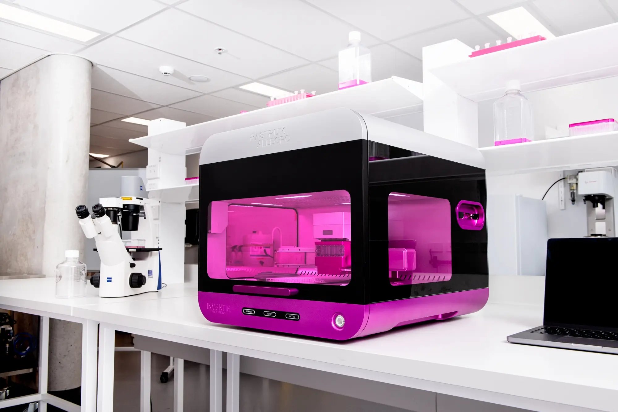
The RASTRUM™ platform helps researchers move from traditional 2D culture to scalable 3D cell culture and 3D cell models that better represent cell behavior, matrix interaction, and disease biology.
Built for drug discovery, disease modeling, and therapeutic research, RASTRUM brings scalable model generation, tunable matrices, defined architectures, guided software, and downstream analysis compatibility into one workflow.
 Scalable 3D cell model generation
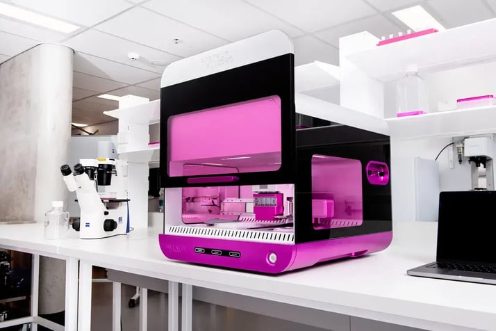
RASTRUM Allegro enables scalable generation of 3D cell models in standard plate formats, helping researchers move from manual setup toward scalable, repeatable workflows.
Scalable 3D cell model generation
RASTRUM Allegro enables scalable generation of 3D cell models in standard plate formats, helping researchers move from manual setup toward scalable, repeatable workflows.
 Tunable cell culture matrices
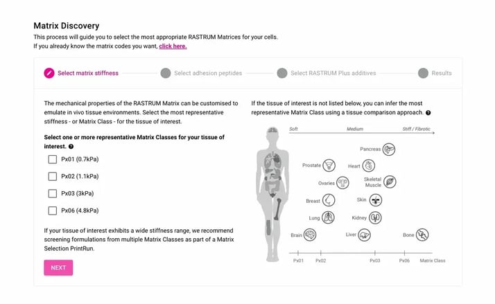
RASTRUM Matrices are xeno-free hydrogels designed to support defined 3D environments. Researchers can select matrix properties such as stiffness, adhesion cues, ECM components, and remodeling behavior based on their model requirements.
Tunable cell culture matrices
RASTRUM Matrices are xeno-free hydrogels designed to support defined 3D environments. Researchers can select matrix properties such as stiffness, adhesion cues, ECM components, and remodeling behavior based on their model requirements.
 Defined cell model architectures
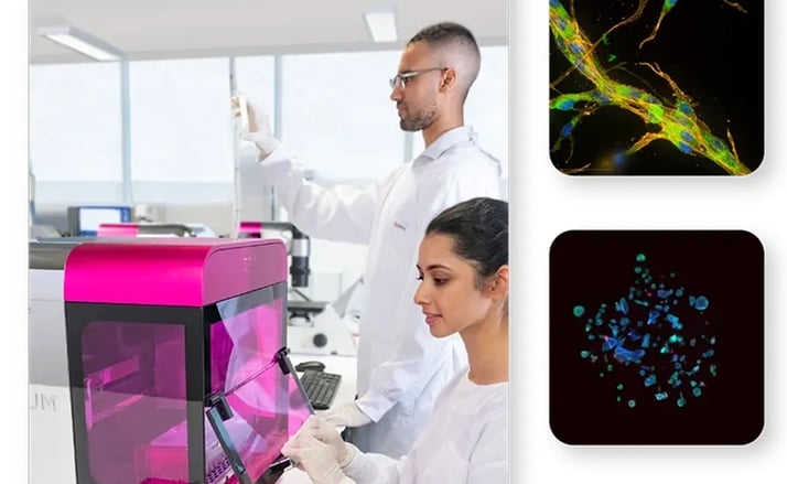
RASTRUM Cell Model Architectures help researchers choose the model format that fits the experiment, including formats for imaging, screening, tissue recovery, spatially defined direct or indirect co-culture, migration, invasion, and other applications.
Defined cell model architectures
RASTRUM Cell Model Architectures help researchers choose the model format that fits the experiment, including formats for imaging, screening, tissue recovery, spatially defined direct or indirect co-culture, migration, invasion, and other applications.
 Guided software workflows
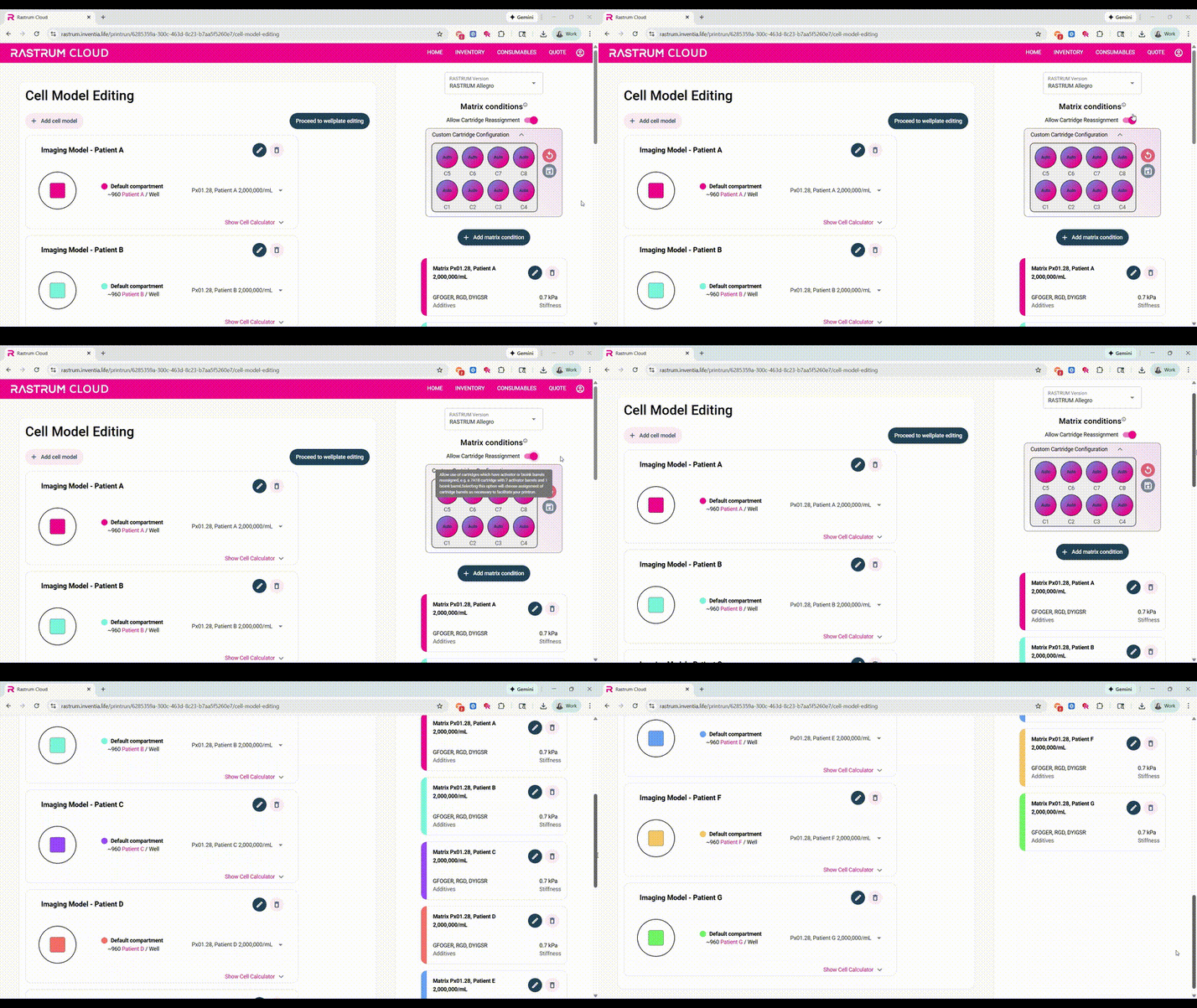
RASTRUM Cloud supports experimental planning, matrix selection, cell density optimization, PrintRun design, protocol generation, and ordering, helping teams standardize setup and reduce workflow ambiguity.
Guided software workflows
RASTRUM Cloud supports experimental planning, matrix selection, cell density optimization, PrintRun design, protocol generation, and ordering, helping teams standardize setup and reduce workflow ambiguity.
 Multiple ways to start
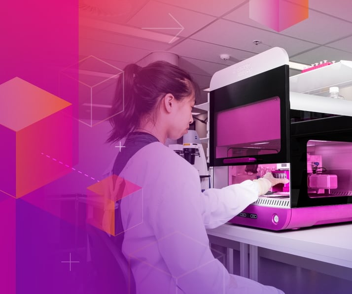
Teams can start with flexible model development, predefined Validated Solutions, or Inventia-supported Discovery Services depending on how defined the biology, workflow, and readouts already are.
Multiple ways to start
Teams can start with flexible model development, predefined Validated Solutions, or Inventia-supported Discovery Services depending on how defined the biology, workflow, and readouts already are.
 Resources
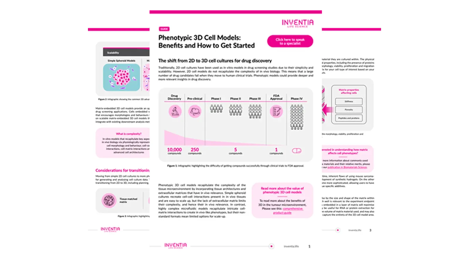
Access application notes, protocols, and publications to enhance your 3D cell culture research with RASTRUM™.
Resources
Access application notes, protocols, and publications to enhance your 3D cell culture research with RASTRUM™.
Cells
Workflows may be designed around immortalized cell lines, primary cells, patient-derived cells, iPSC-derived cells, stromal cells, immune cells, endothelial cells, fibroblasts, or multicellular co-cultures. The appropriate cell system depends on the biological question, model architecture, matrix conditions, culture requirements, and readouts.
Matrix
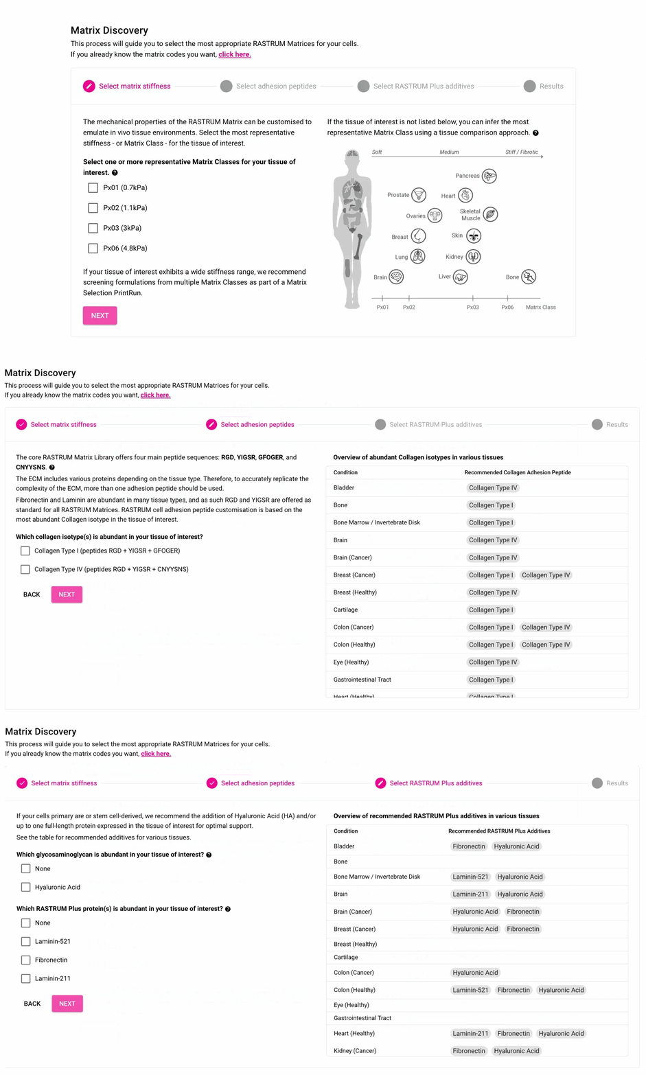
Matrix choice can influence cell viability, morphology, signaling, migration, transport, recovery, and therapeutic response. RASTRUM Matrices help researchers select a defined environment instead of relying only on manual or undefined matrix workflows.
Architecture
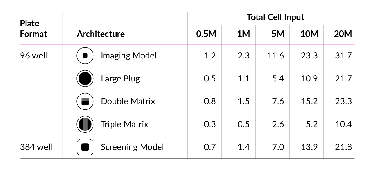
Model architecture determines how cell models are positioned, imaged, measured, recovered, or scaled. Researchers can select architectures based on the application, such as imaging, -omics analysis, high-throughput screening, migration or invasion studies, tissue co-culture, or model recovery.
Readouts
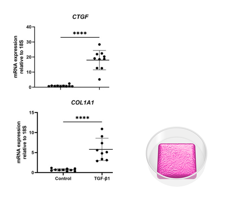
RASTRUM-generated models can be designed around downstream readouts such as 3D cell imaging, viability, morphology, high-content imaging, drug response, molecular analysis, and cell recovery workflows, with readout selection guided by the model format and study design.
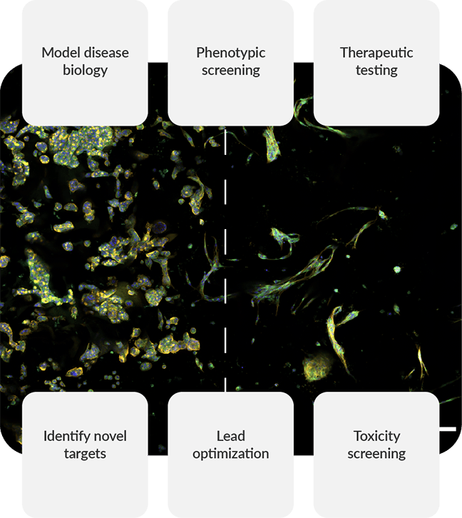
Drug discovery and screening
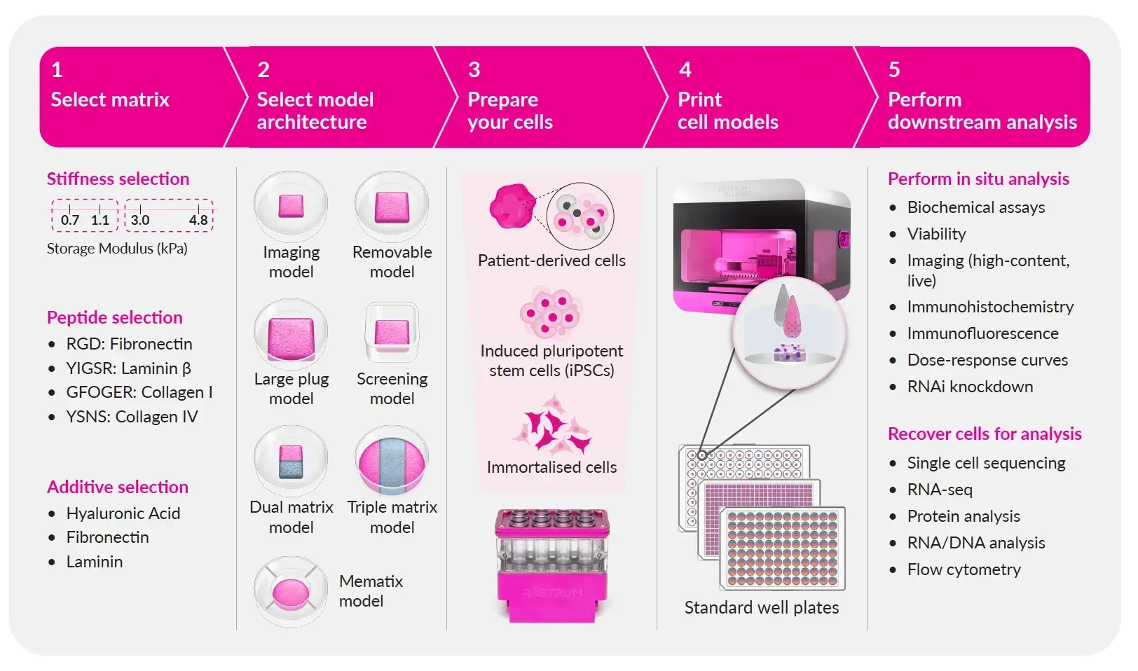
Generate 3D cell models for drug response, lead validation, mechanism studies, and screening. RASTRUM helps teams move from exploratory 3D culture into high-throughput cell culture workflows where reproducibility and scale matter.
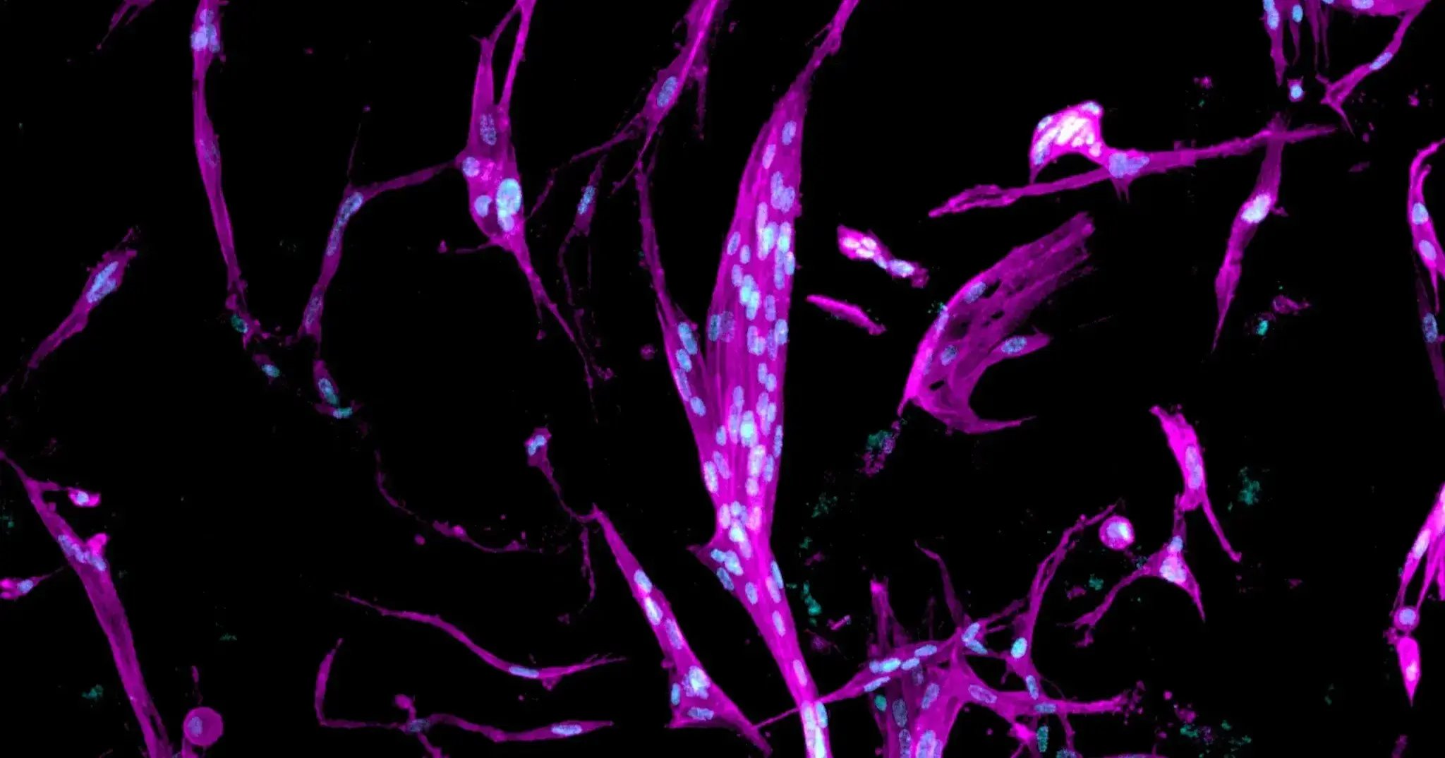
Disease modeling
RASTRUM can support disease model development across cancer, fibrosis, neurodegenerative disease, and other research areas, with project scope defined by the cell source, matrix requirements, architecture, and study goals.
.png?width=1670&height=956&name=image%20(30).png)
Tissue Models
RASTRUM workflows can support tissue model development (including liver, brain, heart, and placenta) using selected cell systems, defined matrices, model architectures, and downstream readouts aligned to the study question.
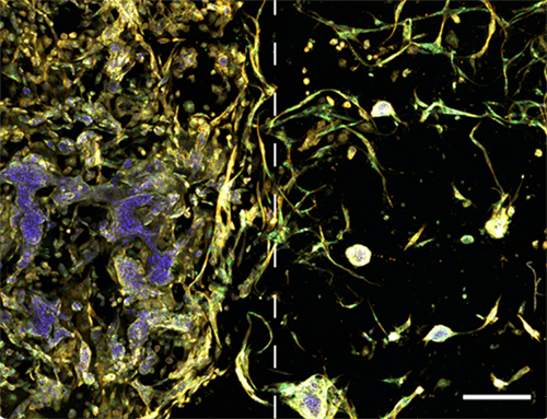
Cancer and tumor microenvironment research
Develop 3D cancer models and tumor microenvironment workflows to study tumor-stroma interaction, immune-cell behavior, therapeutic response, migration, invasion, and patient-derived tumor biology.
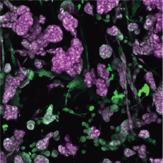
Fibroblast and stromal biology
Use 3D culture to study fibroblast activation, stromal signaling, matrix remodeling, and fibrosis-associated biology. RASTRUM workflows can support fibroblast cell models and more complex stromal co-cultures where appropriate.
Patient-derived and iPSC-derived models
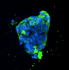
RASTRUM can support 3D cell culture workflows designed around patient-derived or iPSC-derived cells, with model conditions selected for the cell source, sample availability, and readouts.
Imaging and high-content analysis
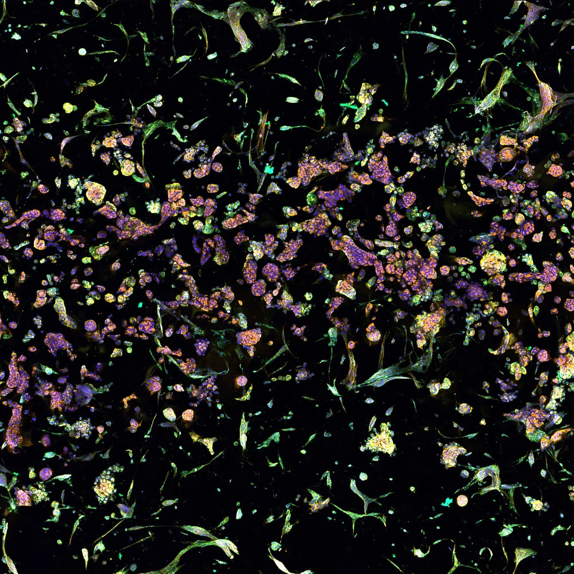
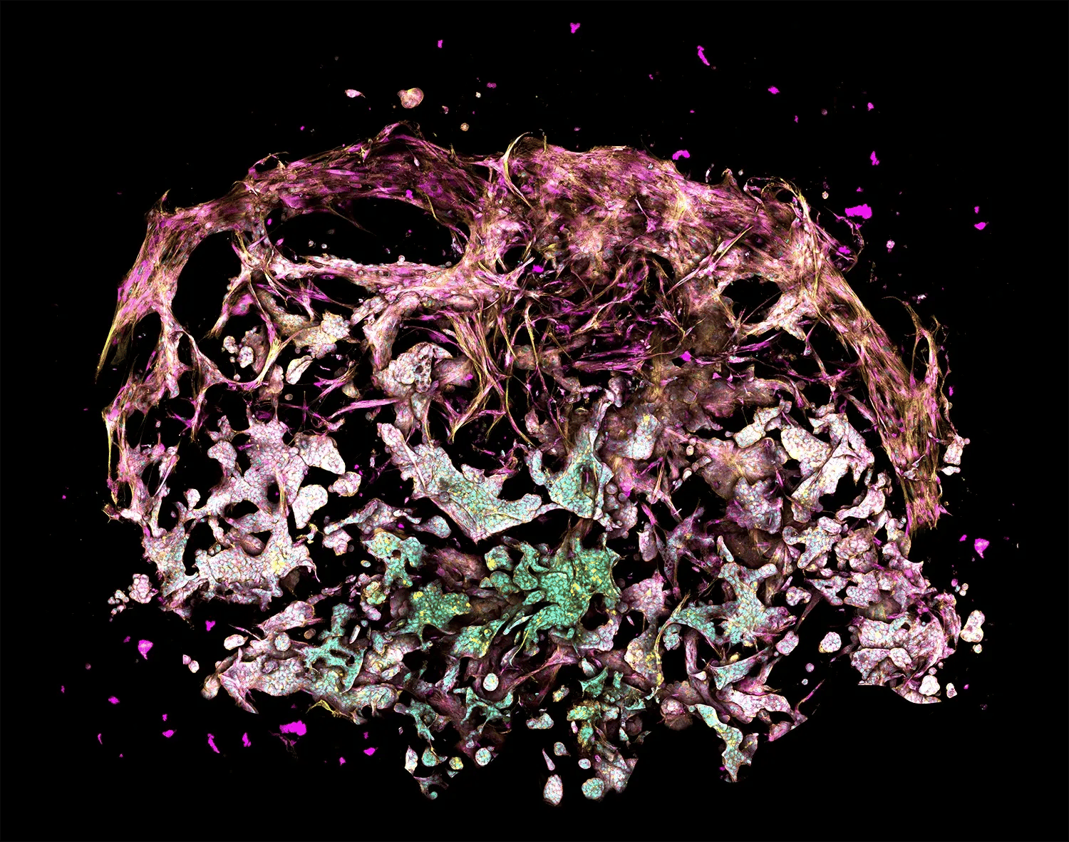
3D cell imaging and 3D cell analysis work best when models are built with consistent architecture and defined matrix conditions, since variability in either can make results harder to interpret or compare across experiments. RASTRUM's standardized, plate-based format is designed to integrate with automated imaging and high-content analysis pipelines, supporting everything from single-cell morphology to multicellular structure and functional readouts.
RASTRUM Allegro
High-throughput 3D cell culture generation for teams that need scale, consistency, and standard plate-based workflows.
 RASTRUM
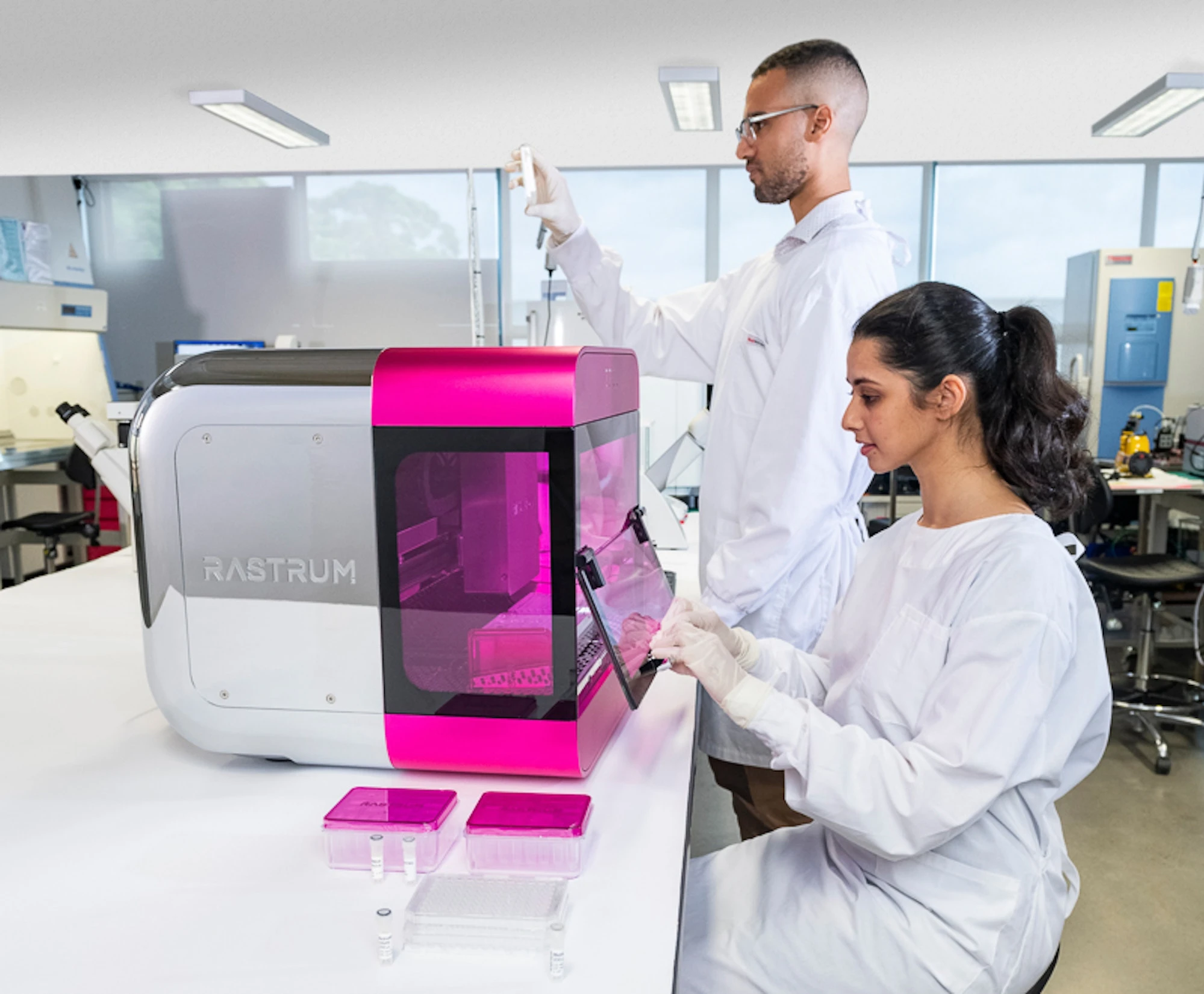
Create precise, matrix-embedded 3D cell models with controlled model design and workflow flexibility.
RASTRUM Cloud
Plan PrintRuns, select model conditions, generate protocols, and support reproducible workflow setup.
RASTRUM
Create precise, matrix-embedded 3D cell models with controlled model design and workflow flexibility.
RASTRUM Cloud
Plan PrintRuns, select model conditions, generate protocols, and support reproducible workflow setup.
 Cell Model Architectures
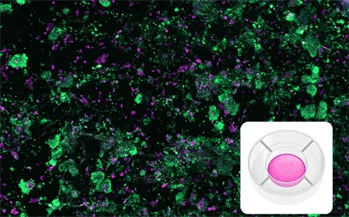
Choose model formats based on what you need to image, screen, recover, interact (directly or indirectly), or scale.
Matrices
Use tunable, xeno-free hydrogels to define the physical and biochemical environment around your cells.
Cell Model Architectures
Choose model formats based on what you need to image, screen, recover, interact (directly or indirectly), or scale.
Matrices
Use tunable, xeno-free hydrogels to define the physical and biochemical environment around your cells.
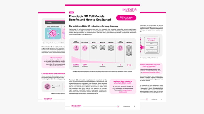 Resources
Access application notes, protocols, publications, webinars, and data packs to support your 3D cell culture research.
I am new to 3D cell culture
Start with educational resources that explain where 3D cell culture models fit, how they differ from 2D systems, and what to consider before choosing a platform.
Resources
Access application notes, protocols, publications, webinars, and data packs to support your 3D cell culture research.
I am new to 3D cell culture
Start with educational resources that explain where 3D cell culture models fit, how they differ from 2D systems, and what to consider before choosing a platform.
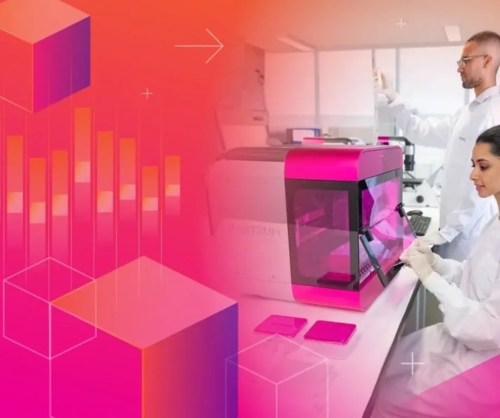 I need to build or optimize a 3D cell model
Explore matrix, architecture, cell density, readout, and workflow choices using Discovery Mode or guided scientific support.
I need to build or optimize a 3D cell model
Explore matrix, architecture, cell density, readout, and workflow choices using Discovery Mode or guided scientific support.
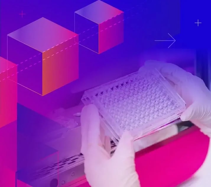 I need a predefined workflow
Start from a Validated Solution when an available context of use matches your biology, readouts, and study goals.
I need a predefined workflow
Start from a Validated Solution when an available context of use matches your biology, readouts, and study goals.
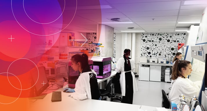 I need a custom 3D disease model
Work with Inventia scientists to assess feasibility, develop model conditions, run studies, generate data, or support transfer of a RASTRUM-based workflow.
I need a custom 3D disease model
Work with Inventia scientists to assess feasibility, develop model conditions, run studies, generate data, or support transfer of a RASTRUM-based workflow.