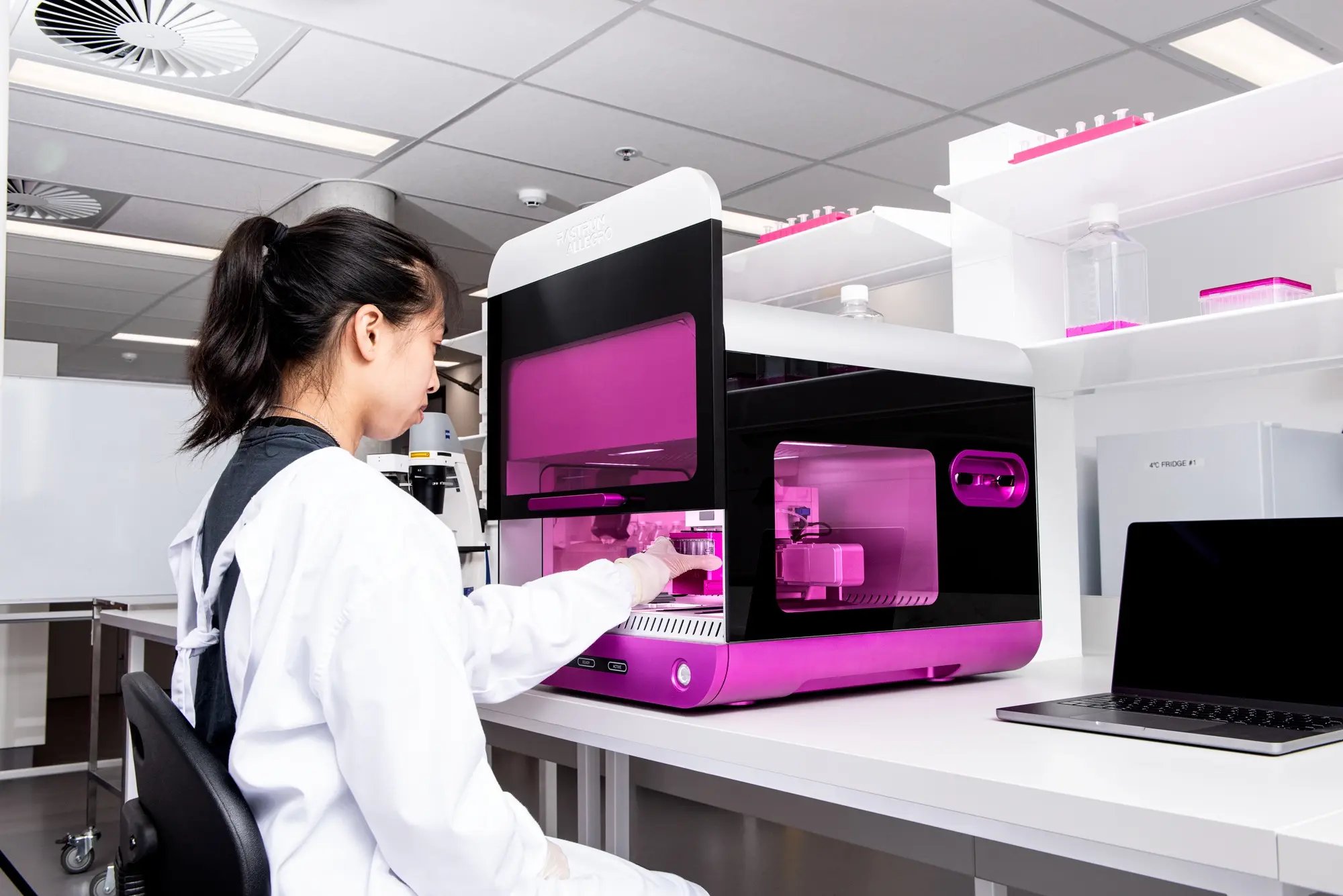
3D disease modeling built around your research question
Every research question places different demands on a 3D model. The right model must capture the cell-cell and cell-matrix interactions that influence the result, while remaining reproducible and compatible with the assays your team already uses.
RASTRUM™ provides a controlled, plate-based way to build matrix-embedded 3D cell models with defined cellular context, matrix conditions, and architecture. Start with your own model concept, adapt an established workflow, or work with Inventia scientists to develop one around your study.
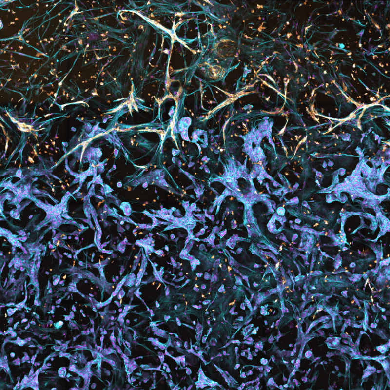
The behavior of cancer cells can change when they are cultured with fibroblasts, immune cells, endothelial cells, or under different matrix conditions. Those influences can affect growth, treatment response, migration, and invasion, and they are easy to miss in a tumor-cell monoculture.
RASTRUM enables integrated or spatially defined co-culture models in tunable matrices, so researchers can isolate the contribution of tumor, stromal, immune, and matrix components. Use these models to test whether a response persists when additional biological context is introduced.
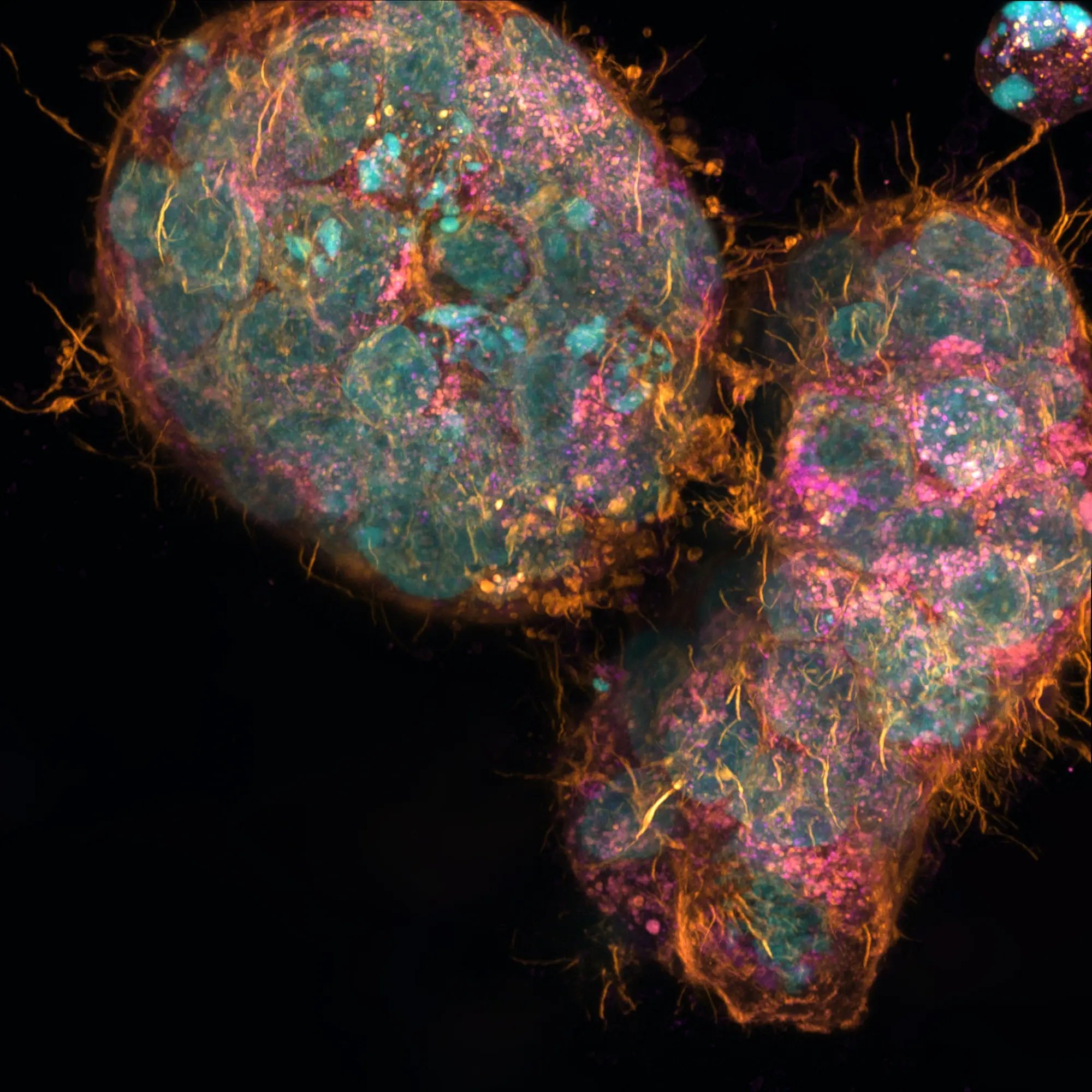
A useful toxicology model has to do more than remain viable at a single endpoint. It must support the exposure schedule, comparison set, and readouts needed to investigate a safety signal or rank compounds.
RASTRUM provides a repeatable, plate-based format for 3D toxicology studies. In established liver workflows, primary human hepatocyte models can be evaluated under acute, chronic, or repeat-dose exposure designs using viability, imaging, and molecular readouts. Similar development approaches may be applied to other toxicology questions in other cell systems using additional assays.
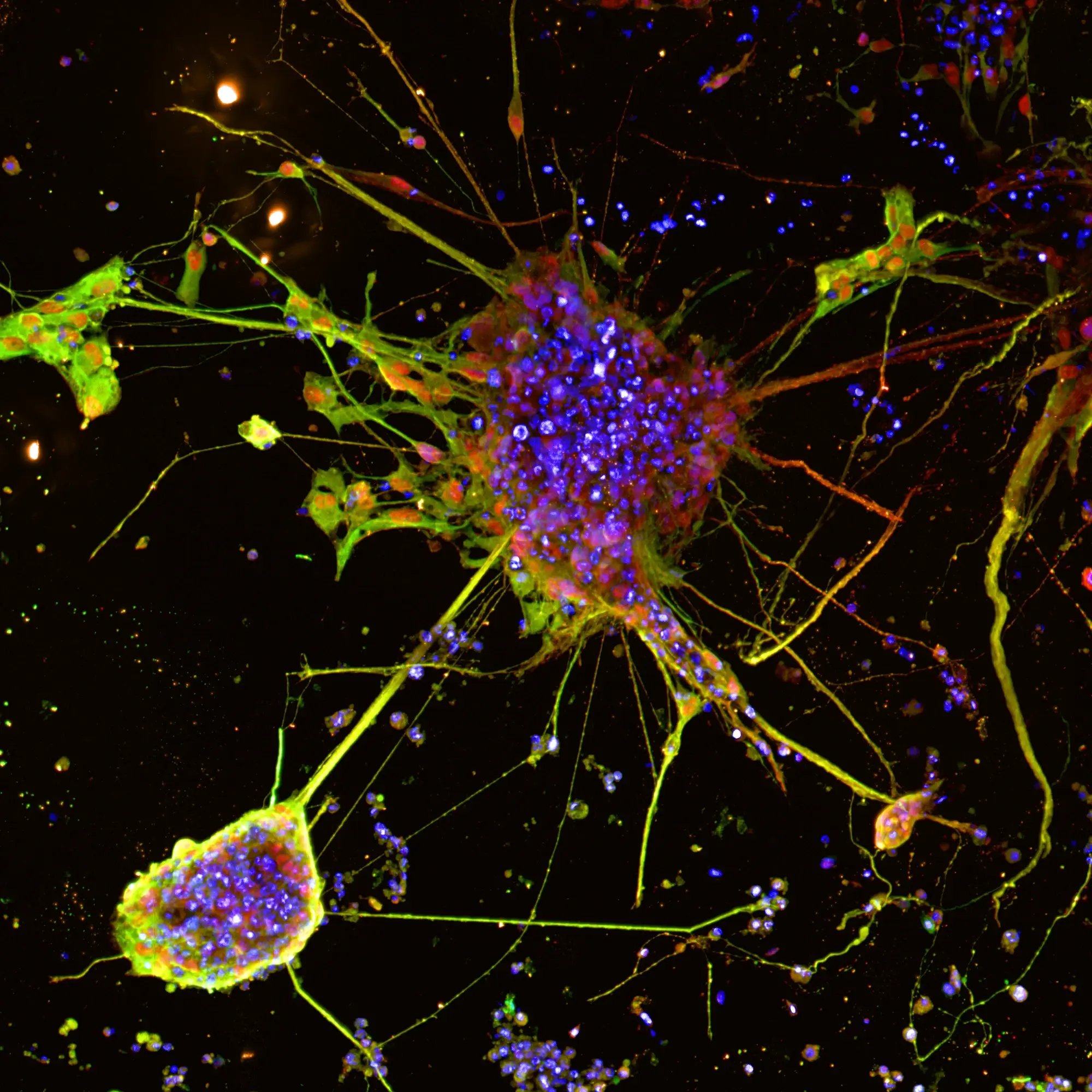
iPSC-derived neural models offer human cell context, but they can be difficult to produce with the consistency and throughput needed for comparative studies. The experimental challenge is to preserve multicellular interactions and structural phenotypes while keeping model generation and analysis practical.
RASTRUM has been used to generate iPSC-derived neuron-astrocyte co-cultures in 96- and 384-well formats. These models showed mature neural markers and neurite and astrocyte projections, and were compatible with cell dyes, immunostaining, and neurite outgrowth assays.
A spatially defined RASTRUM forebrain cortex model has also been used to quantify neurite extension across a hydrogel bridge. In an Alzheimer’s disease study, APP-mutant cultures showed impaired neurite and synapse formation in 3D relative to controls, a difference that was not reproduced in matched 2D cultures.
Together, these workflows demonstrate how neural and neurodegenerative disease research can move beyond cell survival to questions of morphology, connectivity, and disease-associated phenotype.
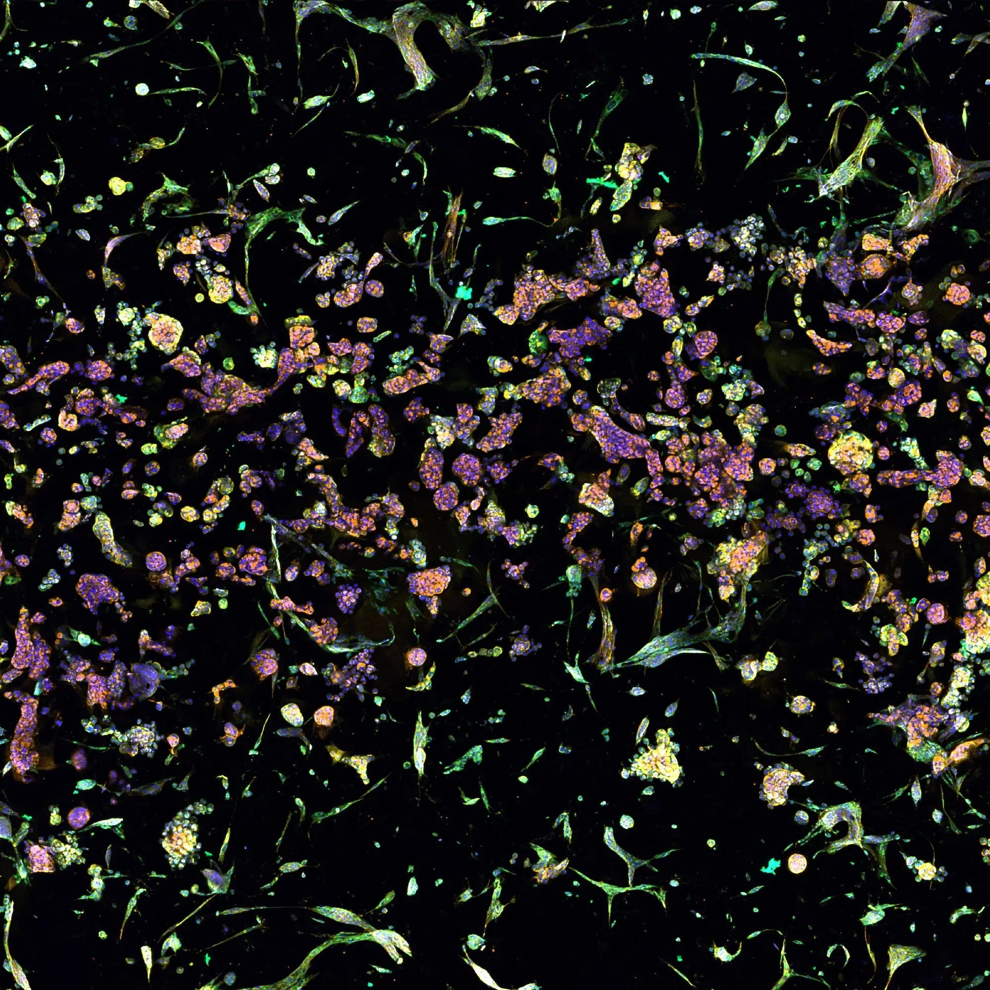
Fibroblast activation state can alter collagen production, stromal signaling, and the behavior of neighboring cells. Yet fibroblasts grown on stiff 2D plastic may become activated before the experiment begins, making it harder to separate a controlled biological stimulus from culture-induced background.
RASTRUM matrices provide a defined 3D environment for establishing a fibroblast model with maintained activation state, then introducing a modulator of fibroblast activity under controlled conditions. In Inventia’s Fibroblast Activation Validated Solution, TGF-β stimulation of normal human lung fibroblasts increased collagen I and COL1A1 expression, providing a measurable activation response.
Use this workflow as a starting point for studies of fibroblast activation, stromal biology, collagen deposition, tumor-stroma interactions, and compounds intended to modulate the activated phenotype.
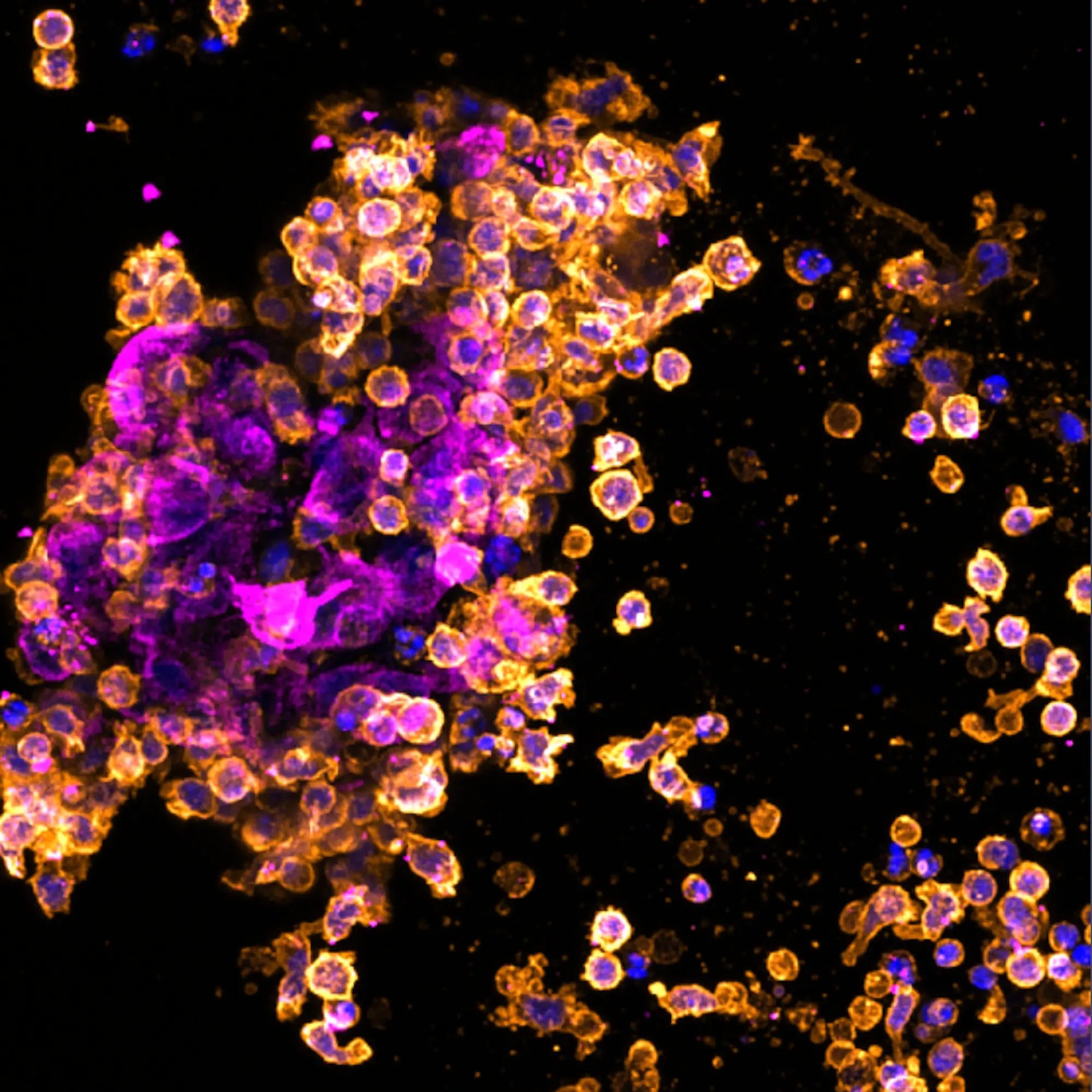
Endpoint tumor killing is only one part of an immuno-oncology experiment. The harder question is whether immune cells can reach tumor cells, engage with them, and mount a cytotoxic response, and how the matrix or stromal context changes each step.
RASTRUM enables direct tumor-immune co-cultures and infiltration models in defined 3D matrices. Researchers can measure immune-cell infiltration and co-localization alongside tumor growth and cytotoxicity, rather than relying on a single endpoint.
In a RASTRUM model using A549 lung cancer cells, activated peripheral blood mononuclear cells inhibited tumor growth in direct co-culture. A related model demonstrated cytotoxic T lymphocyte infiltration and showed that matrix stiffness modulated T cell motility and cytotoxicity. These models can help researchers examine whether a response is determined by immune-cell access, the surrounding matrix, or tumor-immune interaction.
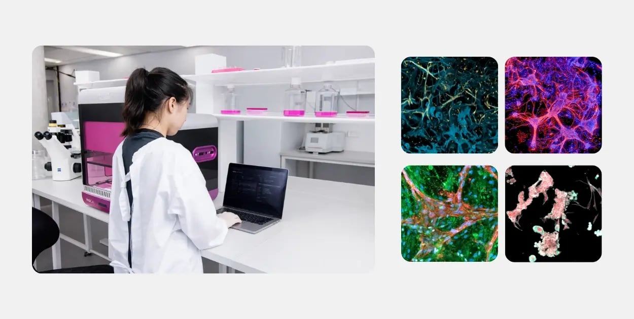
Is RASTRUM a fit for your model?
Your study may span more than one of these areas, or start with a cell system that has not yet been shown here. Share the biological question, cell source, experimental scale, and intended readout with our team. We can help determine whether RASTRUM is a practical fit and whether your best starting point is flexible in-house development, a validated workflow, or hands-on support from Discovery Services.
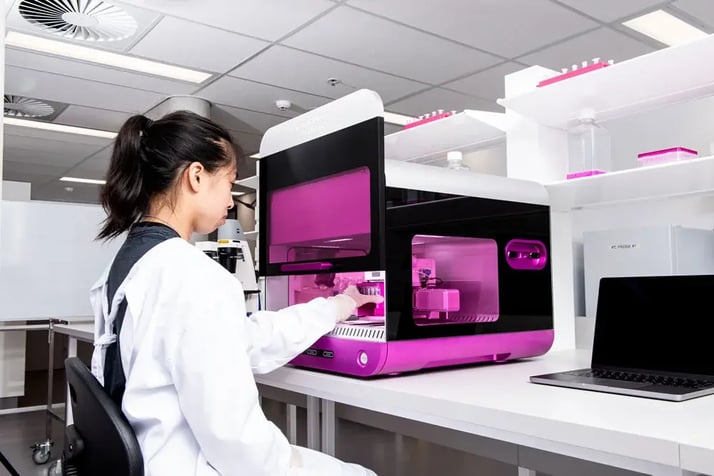 RASTRUM™ Allegro
Generate reproducible, scalable 3D cell models in standard well-plate formats.
RASTRUM™ Allegro
Generate reproducible, scalable 3D cell models in standard well-plate formats.
 RASTRUM™ Cell Model Architectures
Choose the physical model configuration that fits what you need to model, measure, or scale.
RASTRUM™ Cell Model Architectures
Choose the physical model configuration that fits what you need to model, measure, or scale.
 RASTRUM™ Matrices
Create defined 3D microenvironments with tunable, xeno-free hydrogels.
RASTRUM™ Matrices
Create defined 3D microenvironments with tunable, xeno-free hydrogels.