
PDAC Validated Solution
A defined 3D PDAC tumor-CAF co-culture workflow for studying tumor-stroma interactions, CAF-mediated chemoprotection, and therapeutic response.
The PDAC Validated Solution combines pancreatic cancer cells with primary PDAC CAFs in a defined 3D matrix environment, enabling researchers to evaluate how stromal biology alters drug response across PANC-1 and AsPC-1 model contexts.
-1.webp?width=714) Tumor-stroma crosstalk
Study interactions between PDAC cancer cells and primary CAFs in a defined 3D matrix environment.
Tumor-stroma crosstalk
Study interactions between PDAC cancer cells and primary CAFs in a defined 3D matrix environment.
 CAF-mediated chemoprotection
Evaluate how stromal biology alters therapeutic response and contributes to drug resistance.
CAF-mediated chemoprotection
Evaluate how stromal biology alters therapeutic response and contributes to drug resistance.
 Genotype-dependent signaling
Compare PANC-1 and AsPC-1 tumor-stroma contexts to investigate differential pathway activation.
Genotype-dependent signaling
Compare PANC-1 and AsPC-1 tumor-stroma contexts to investigate differential pathway activation.
 Combination therapy evaluation
Test strategies designed to counteract stromal-mediated resistance or remain effective in CAF-rich contexts.
Combination therapy evaluation
Test strategies designed to counteract stromal-mediated resistance or remain effective in CAF-rich contexts.

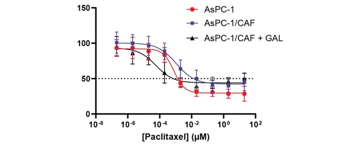
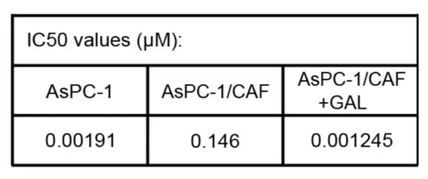
Proof point: CAF co-culture alters therapeutic response in 3D PDAC models
In co-culture models, CAF interactions increased resistance to paclitaxel relative to monoculture conditions. Addition of galunisertib reversed the chemoprotective effect, supporting use of the workflow for evaluating combination strategies targeting stromal-mediated resistance.

Context of use
This validated workflow is designed for researchers who need a reproducible 3D PDAC tumor-stroma model for therapeutic response studies in a defined context of use.
- Evaluate therapeutic efficacy in the presence of tumor-stroma crosstalk
- Investigate stromal contributions to tumor progression and drug resistance
- Identify targetable resistance mechanisms in the tumor microenvironment
- Screen pathway-targeted therapies and combination treatments
Image depicts immunofluorescence assessment of cancer cell–CAF interactions in RASTRUM optimized models. Positive immunofluorescence staining for pan cytokeratin (cancer cell marker, green) alongside phalloidin staining for actin filaments (red) after 7 days in culture. (A) PANC-1/CAF and (B) AsPC-1/CAF co-cultures.

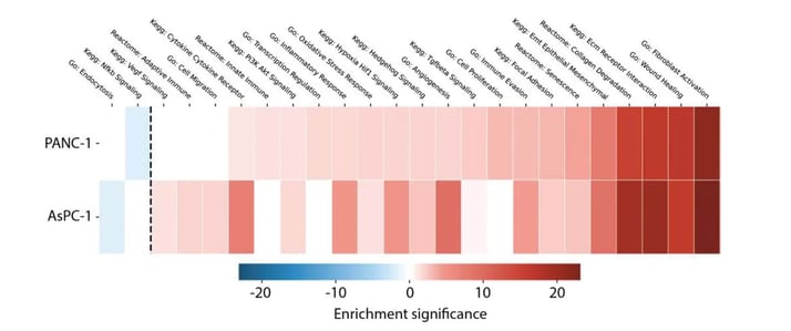
Molecular characterization
RNA-seq reveals pathway activation associated with tumor-stroma signaling
Differential gene expression analysis highlights pathway enrichment related to fibroblast activation, pro-tumourigenic processes, and therapeutic resistance. Distinct pathway profiles between PANC-1 and AsPC-1 co-cultures support the use of this workflow in biologically rich PDAC contexts.
Functional validation
Tumor-stroma interactions alter therapeutic response
Co-culture models demonstrate increased resistance to paclitaxel relative to monoculture conditions, consistent with stromal-mediated chemoprotective effects observed in PDAC. Addition of the TGFBR1 inhibitor galunisertib reverses the chemoprotective effect in co-cultures, supporting use of this model for combination therapy evaluation.
 Download the Data Pack
Best for evaluating whether this defined 3D PDAC tumor-CAF co-culture workflow fits your research question.
Download the Data Pack
Best for evaluating whether this defined 3D PDAC tumor-CAF co-culture workflow fits your research question. Review supporting data for model setup, CAF phenotype, tumor-CAF interactions, and therapeutic response in defined PANC-1/CAF and AsPC-1/CAF contexts.
Download the Data Pack
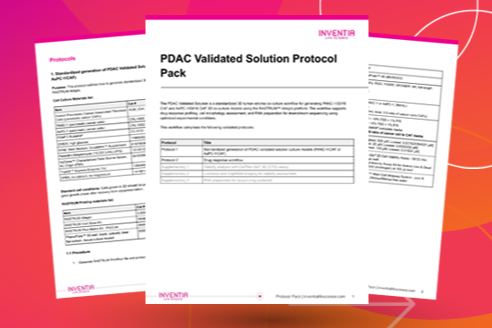 Get the Protocol Pack
Best for teams preparing to run or evaluate implementation of the workflow.
Get the Protocol Pack
Best for teams preparing to run or evaluate implementation of the workflow.Includes protocols for model generation, drug-response testing, viability analysis, imaging, and optional RNA preparation for sequencing.
Get the Protocol Pack
 Need a different PDAC context?
Need a different cell source, matrix condition, co-culture, readout, or context of use?
Need a different PDAC context?
Need a different cell source, matrix condition, co-culture, readout, or context of use?Talk with Inventia scientists about whether Discovery Services can help assess or develop a custom PDAC model.
Discuss a custom PDAC model
 Browse Validated Solutions
Predefined blueprints for reproducible 3D data in a defined context of use
Browse Validated Solutions
Predefined blueprints for reproducible 3D data in a defined context of use
 Explore Discovery Mode
Define and refine 3D cell models to match your biology and readouts
Explore Discovery Mode
Define and refine 3D cell models to match your biology and readouts
 Connect with Discovery Services
Expert support to optimize, validate, and integrate 3D models into discovery workflows
Connect with Discovery Services
Expert support to optimize, validate, and integrate 3D models into discovery workflows