
Matrix mechanics and culture conditions can influence fibroblast state before treatment begins, complicating the interpretation of stromal biology and drug response.
Fibroblast activation is often measured as an experimental outcome. Yet the culture environment may have already influenced fibroblast contractility, matrix production, and signaling before the first treatment is added.
For discovery and translational teams, this creates an interpretation problem. When an activation-associated marker changes after treatment, how much of the response came from the perturbation? How much was established by the matrix, substrate mechanics, cell history, or culture conditions?
The answer can affect assay window, mechanism-of-action studies, compound ranking, and confidence in tumor-stroma data. A model intended to measure fibroblast activation needs a well-characterized starting state so that treatment-induced biology can be distinguished from biology introduced by the workflow.
The matrix can shape fibroblast state before treatment begins
Fibroblasts respond closely to the physical and biochemical properties of their surroundings.
Matrix stiffness can influence contractility, proliferation, extracellular matrix production, and response to biochemical signals such as transforming growth factor beta, or TGF-β. A rigid culture surface may push cells toward activation-associated behavior before an experimental stimulus is introduced. An undefined matrix can add another source of uncertainty if its mechanical or biochemical properties vary between experiments.
For compound studies, an elevated baseline can reduce the dynamic range of the assay. It may also become harder to determine whether a candidate is changing fibroblast state, compensating for a culture-induced phenotype, or affecting cell health more broadly.
This makes matrix selection part of the assay design.
RASTRUM™ Matrices are synthetic PEG-based environments that can be configured using different stiffnesses, adhesion peptides, full-length extracellular matrix molecules, and matrix metalloproteinase-sensitive components. These defined properties allow scientists to investigate selected mechanical and biochemical cues without relying on an undefined matrix composition.
The formulation still needs to fit the fibroblast source, tissue context, and experimental question. A condition suited to one cell population or activation mechanism may not be appropriate for another.
Fibroblast activation spans multiple functional states
Fibroblast activation is not represented by one universal phenotype.
A contractile myofibroblast state is commonly associated with changes in actin organization, collagen production, contractility, and extracellular matrix remodeling. Other activated states may be characterized more strongly by inflammatory signaling, cytokine production, altered metabolism, or interactions with neighboring cells.
This heterogeneity is especially relevant among cancer-associated fibroblasts, or CAFs. Studies in pancreatic ductal adenocarcinoma have identified spatially and functionally distinct myofibroblastic and inflammatory CAF populations. Tumor-derived TGF-β and interleukin-1 signaling can help shape these states, demonstrating that local context influences both fibroblast identity and function.
No single marker defines fibroblast activation across every tissue and experimental setting. Alpha-smooth muscle actin may be highly informative in one context, while collagen expression, cytokine secretion, morphology, contractility, or pathway activity may be more useful in another.
The intended context of use should determine the marker panel. Scientists need to define the fibroblast state they expect to observe and select readouts capable of distinguishing that state from changes in viability, proliferation, or general cellular stress.
The same principle applies across related areas of stromal biology. A wound healing model may examine transient activation, migration, matrix deposition, and resolution after a defined stimulus. A fibrosis model may focus on sustained myofibroblast activity and continued extracellular matrix production. Within the tumor stroma, the question may involve CAF state, tumor-fibroblast signaling, immune-cell access, or the effect of stromal biology on therapeutic response.
These applications share biological features, but each requires its own context of use, controls, and readouts.
Why fibroblast state matters in tumor-stroma drug studies
Fibroblasts can have a substantial influence on the extracellular matrix and signaling environment surrounding a tumor.
Activated fibroblasts may affect matrix organization, tissue mechanics, soluble signaling, tumor-cell phenotype, and immune-cell behavior. CAF populations can also respond differently to the same perturbation, depending on tumor type, cellular state, spatial location, and the surrounding microenvironment.
The Fibroblast Activation Data Sheet notes that fibroblast activation and the resulting extracellular matrix remodeling can modulate tumor-cell biology and reduce the efficacy of drug treatments, including immunotherapies.
Those effects create several possible sources for a drug-response signal. A treatment may act directly on the cancer cells, change fibroblast state, alter signaling between the two populations, or produce effects across several components of the model.
For discovery teams, the experimental questions may include:
- Does the candidate alter fibroblast activation or matrix production?
- Does tumor-cell response change in the presence of fibroblasts?
- Are direct contact or paracrine signals influencing the response?
- Which cell population accounts for the observed effect?
Model architecture can help separate these mechanisms.
The Fibroblast Activation Data Sheet shows NHLF and A549 lung cancer cells together in an Imaging Model and spatially separated in a Dual Matrix Model designed to support investigation of paracrine signaling. These examples demonstrate how direct and spatially organized cocultures can be used to study different aspects of fibroblast-cancer cell crosstalk.
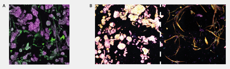 Figure 1. Direct and spatially separated fibroblast-cancer cocultures generated with RASTRUM. NHLF and A549 lung cancer cells were cultured together in an Imaging Model or spatially separated in a Dual Matrix Model to support investigation of paracrine signaling. These examples demonstrate broader coculture capability and are separate from the validated NHLF activation workflow.
The model should be built around the decision the team needs to make. Tumor-only, fibroblast-only, untreated, stimulated, direct coculture, and spatially separated conditions may each contribute a different part of the answer.
The aim is interpretable context: enough biological complexity to represent the stromal question, combined with sufficient control to explain where the resulting signal came from.
For a broader discussion of immune, endothelial, stromal, and spatial components within the tumor microenvironment, see Creating Dynamic Tumor Microenvironments with 3D Cell Models. The present article remains focused on fibroblast activation as an experimental variable.
Designing for interpretable fibroblast-response data
Before a compound study begins, teams need a characterized baseline, readouts suited to the biological question, and a repeatable workflow.
A characterized baseline. Cell source, passage history, previous culture conditions, viability, morphology, and context-relevant activation markers should be documented in the unstimulated condition. A defined activation protocol can then confirm that the cells retain the capacity to respond.
Readouts that resolve the biological question. Viability can indicate that a culture has changed, but imaging, gene-expression analysis, or cell-specific measurements may be needed to determine whether the effect came from fibroblasts, tumor cells, or both populations.
A repeatable workflow. Cell input, matrix conditions, model architecture, treatment timing, and assay execution should remain controlled across wells, plates, and studies. Variation in model production can otherwise resemble biological heterogeneity or pharmacological response.
Together, these elements provide a stable framework for determining whether a perturbation changed fibroblast state or produced broader effects on cell health and model behavior.
A defined 3D starting point for fibroblast activation studies
Inventia’s Fibroblast Activation Validated Solution provides a standardized workflow for generating 3D normal human lung fibroblast, or NHLF, models under defined operating conditions.
The workflow uses NHLF cells at 10 million cells/mL in a defined 1.1 kPa RASTRUM Matrix containing RGD, YIGSR, GFOGER, and hyaluronic acid. Imaging Model and Large Plug Model formats are used depending on the downstream analysis.
Fibroblast activation is induced by adding TGF-β at 20 ng/mL on day 4 post-printing for 24 hours. The response is evaluated through collagen I detection by immunofluorescence and COL1A1 gene-expression analysis by qPCR. Optional protocols support brightfield and live/dead imaging and CellTiter-Glo 3D viability analysis.
In the supporting Data Pack, NHLF cells remained highly viable for at least seven days in the tested RASTRUM Matrix conditions.
Following TGF-β treatment, the models showed increased collagen I staining and increased COL1A1 expression relative to untreated controls. The Data Sheet reports an approximately fourfold increase in COL1A1 mRNA expression following treatment.
The untreated models provide an activatable baseline against which the TGF-β-induced response can be measured. This baseline supports comparison between unstimulated and stimulated conditions within the same defined model environment.
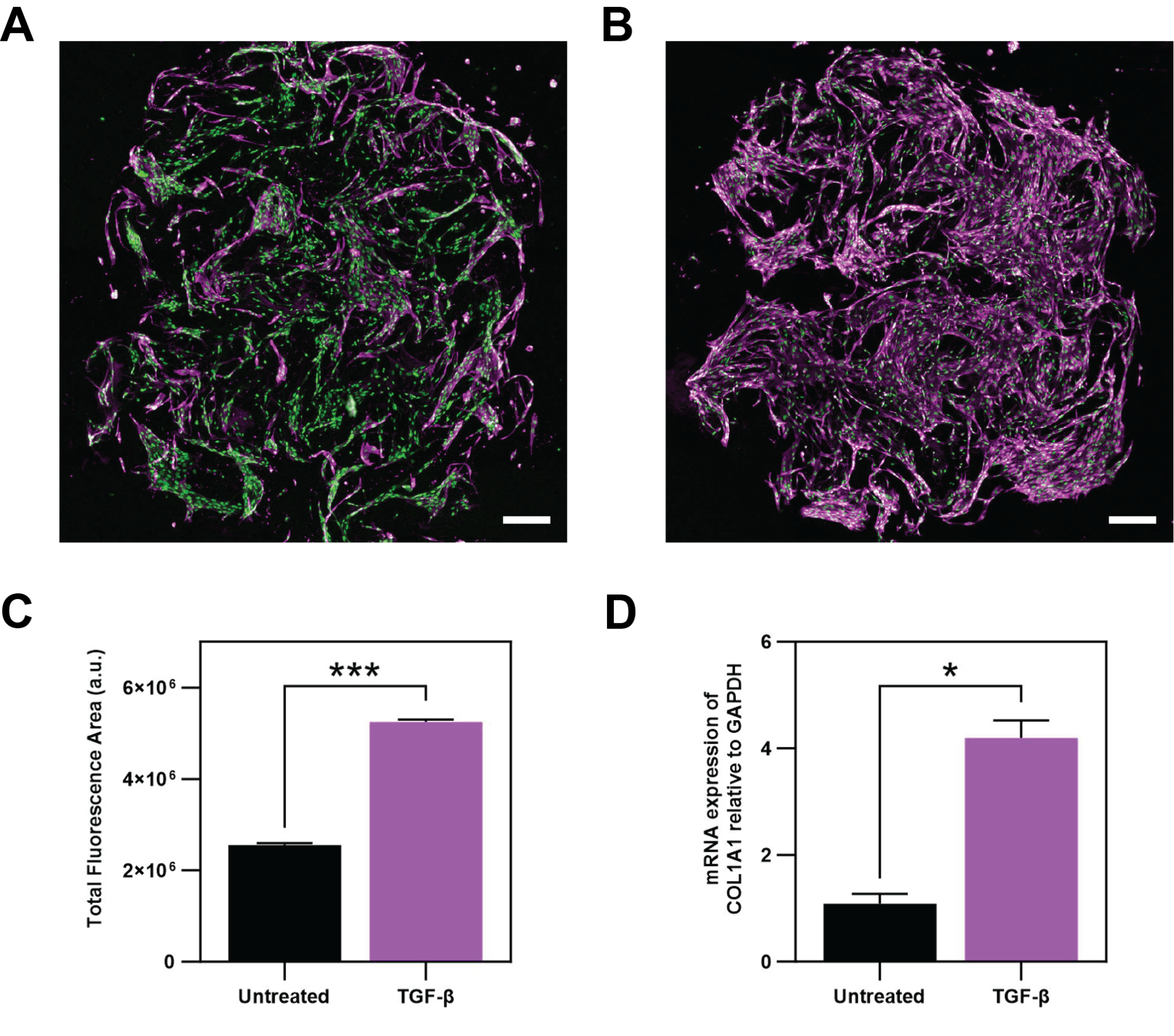 Figure 2. TGF-β-induced fibroblast activation in RASTRUM 3D models. NHLF models treated with TGF-β showed increased collagen I staining and increased COL1A1 expression relative to untreated controls under the validated workflow conditions.
The Protocol Pack includes validated protocols for:
- standardized NHLF model generation
- TGF-β induction of fibroblast activation
- collagen I detection by immunofluorescence
- COL1A1 gene-expression analysis by qPCR
- optional brightfield and live/dead imaging
- optional CellTiter-Glo 3D viability analysis
The workflow is documented with a defined model configuration, treatment schedule, downstream analyses, validated protocols, and supporting example data.
The Data Pack includes representative morphology, viability through at least seven days, collagen I immunofluorescence and image-based quantification, COL1A1 expression, and comparative collagen I imaging in RASTRUM Matrix and basement membrane extract. Under the compared imaging conditions, collagen I staining was visualized more clearly in the RASTRUM Matrix.
Together, these materials provide a documented starting point for studying fibroblast activation and evaluating strategies intended to perturb that response.
The validated context of use is specific to the documented NHLF workflow. Different fibroblast sources, matrices, activation mechanisms, disease settings, or readouts require their own optimization and validation.
From activation assays to more complex stromal biology
The validated core workflow centers on NHLF monoculture and TGF-β-induced activation.
Once a baseline activation assay has been established, teams may extend the model to questions involving tumor-fibroblast interactions, compound response, or additional tissue-specific stromal biology.
The direct and spatially separated NHLF-A549 coculture examples in the Data Sheet illustrate broader model configurations supported by RASTRUM. They are separate from the validated monoculture activation protocols documented in the Protocol Pack.
More complex questions may require different fibroblast sources, tumor or immune cells, or architectures that separate cell populations spatially. In those settings, the same experimental principles remain useful:
- establish the starting state
- define the activation mechanism
- select readouts that resolve cell-specific effects
- document the variables that influence the phenotype
- confirm that the workflow remains reproducible as complexity increases
The model should become more complex when the scientific question requires it. Each added cell type, matrix cue, architecture, and readout should have a defined role in the hypothesis.
Better stromal data starts before treatment
Fibroblast activation should be considered from the beginning of assay design.
Before screening a compound or interpreting tumor-stroma signaling, discovery teams need a clear view of the fibroblast baseline, the cues supplied by the matrix, and the readouts used to define the response.
A controlled starting state creates a clearer connection between perturbation and phenotype. It also provides a stronger foundation for comparing treatments, investigating mechanisms, and deciding which findings warrant further study.
Defining the starting biology reduces background uncertainty and makes treatment-induced changes easier to interpret and act on.
Explore fibroblast and stromal biology with RASTRUM
Learn more about:
- the Fibroblast Activation Validated Solution
- the Fibroblast Activation Data Sheet
- the Fibroblast Activation Data Pack
- the Fibroblast Activation Protocol Pack
- Fibroblast and stromal biology in Inventia’s Areas of Research
- tunable RASTRUM Matrices
- 3D models for Cancer research
- Cell Model Architectures for coculture and spatially organized models
- Creating Dynamic Tumor Microenvironments with 3D Cell Models
- Discovery Services for customized fibroblast and stromal model development
Or connect with an Inventia 3D biology specialist to discuss what your team would need to establish a reproducible 3D cell culture workflow.