PDAC-IO Validated Solution
A defined 3D PDAC tumor-stroma-immune model for immuno-oncology (IO) research
The PDAC-IO Validated Solution is a 3D tri-culture of PANC-1 pancreatic cancer cells, primary cancer-associated fibroblasts (CAFs) and pre-activated human CD8+ T cells, built by adding an immune component to the PDAC Validated Solution. Use the supporting imaging, bulk RNA-seq, and reference-treatment data to assess scientific fit, then use the Protocol Pack to implement the workflow in your lab.
Model at a glance
A defined starting point for more contextual PDAC research
PDAC research often requires more biological context than cancer cells alone can provide. Bringing tumor, stromal and immune components together in a controlled 3D model requires coordinated decisions about cell composition, matrix, immune-cell preparation, timing and readouts.
The PDAC-IO Validated Solution brings those parameters together in a documented model with supporting data, helping teams assess fit and begin from an established configuration rather than developing every element from scratch.
- Disease area: Pancreatic ductal adenocarcinoma (PDAC)
- Model type: 3D tumor-stroma-immune tri-culture
- Cells: PANC-1 cancer cells, primary PDAC CAFs, pre-activated human CD8+ T cells (15,000 per well)
- Matrix: Px03.84, about 3 kPa, with RGD, YIGSR, GFOGER, hyaluronic acid and full-length fibronectin.
- Research focus: Tumor-stroma-immune interactions, CAF-associated immunoregulation, cancer cell-death response to immunomodulators
- Readouts: Cleaved caspase-3 and pan-cytokeratin immunofluorescence
- Reference compounds: Pembrolizumab (anti-PD-1), relatlimab (anti-LAG-3), galunisertib, human IgG4 isotype control
What this workflow helps you investigate
Tumor-stroma-immune interactions
Study cancer cells, CAFs and CD8+ T cells together in a defined 3D matrix
CAF-associated immunoregulation
Study CAF effects on immune cell interactions with cancer cells
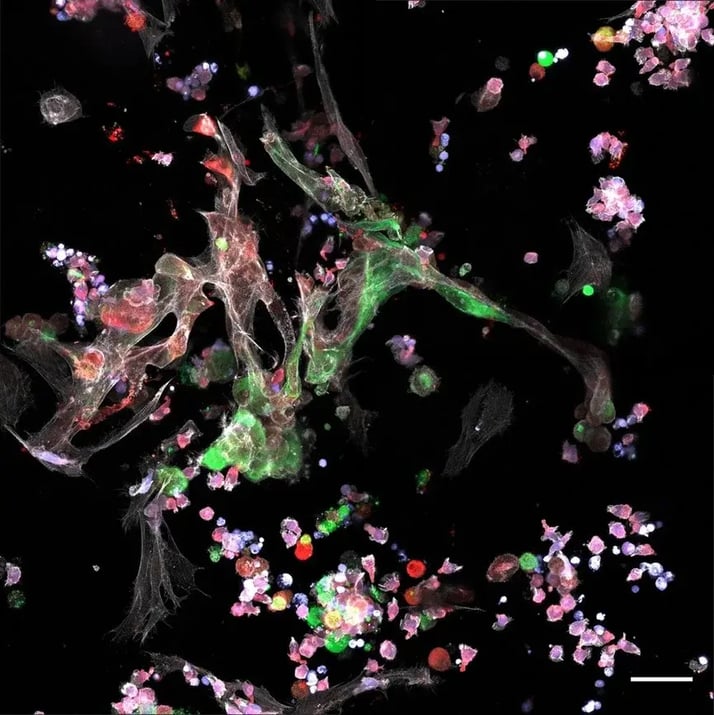
Checkpoint inhibitor response and stroma-targeted combinations
Measure cancer cell death after anti-PD-1, alone or combined with anti-LAG-3 or the TGF-β receptor I kinase inhibitor galunisertib, in a CAF-containing context
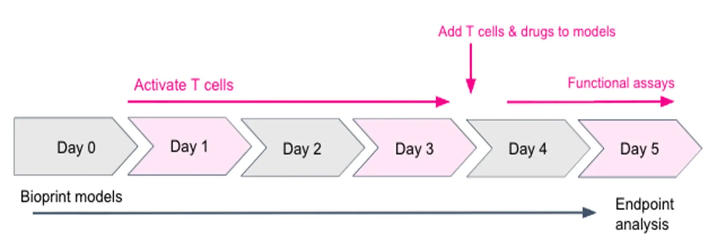
PDAC-IO experimental timeline. Day 0: print PANC-1 and CAF co-culture. Day 1: revive and activate CD8+ T cells (72 hours). Day 4: add CD8+ T cells and treatment (24 hours). Day 5: collect RNA, or fix for immunofluorescence staining.
Context of use
This validated workflow enables researchers to:
- Evaluate immunotherapy efficacy in the presence of tumor-stroma-immune crosstalk
- Investigate how cancer-associated fibroblasts (CAFs) shape tumor signaling and CD8+ T cell function
- Identify targetable mechanisms of stroma-mediated immune suppression within the tumor microenvironment
- Screen immune checkpoint inhibitors and stroma-targeted combination treatments
The PDAC-IO Validated Solution adds an immune component to the PDAC Validated Solution, integrating pancreatic cancer cells, CAFs and CD8+ T cells within a defined 3D matrix. Because the tumor and CAF compartments are established before T cells are added, early immune-driven changes can be measured against a mature, stroma-conditioned baseline.
It is built for teams working on pancreatic cancer immunotherapy and tumor microenvironment biology. For research use only.
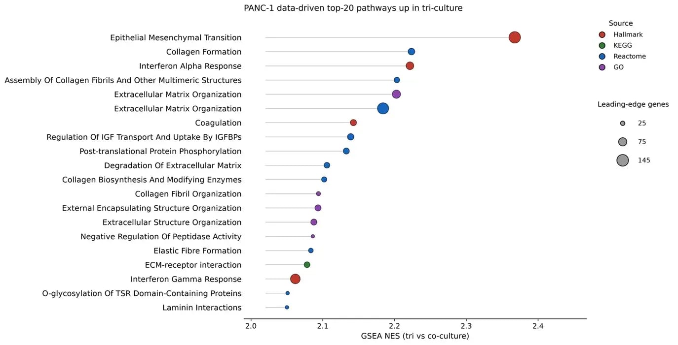
Molecular characterization
Bulk RNA-seq shows CAF-associated changes in pathway activity
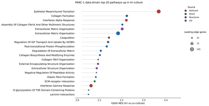
RNA was collected 24 hours after CD8+ T cell addition (day 5 after printing) and analyzed by bulk RNA sequencing. Gene set enrichment analysis compared PANC-1 tri-cultures (PANC-1, CAFs and CD8+ T cells) with co-cultures of PANC-1 and CD8+ T cells. Tri-cultures were enriched for epithelial-mesenchymal transition, collagen formation, extracellular matrix organization, and interferon alpha and gamma response gene sets (top 20 pathways by normalized enrichment score, all FDR < 0.001).
Functional validation
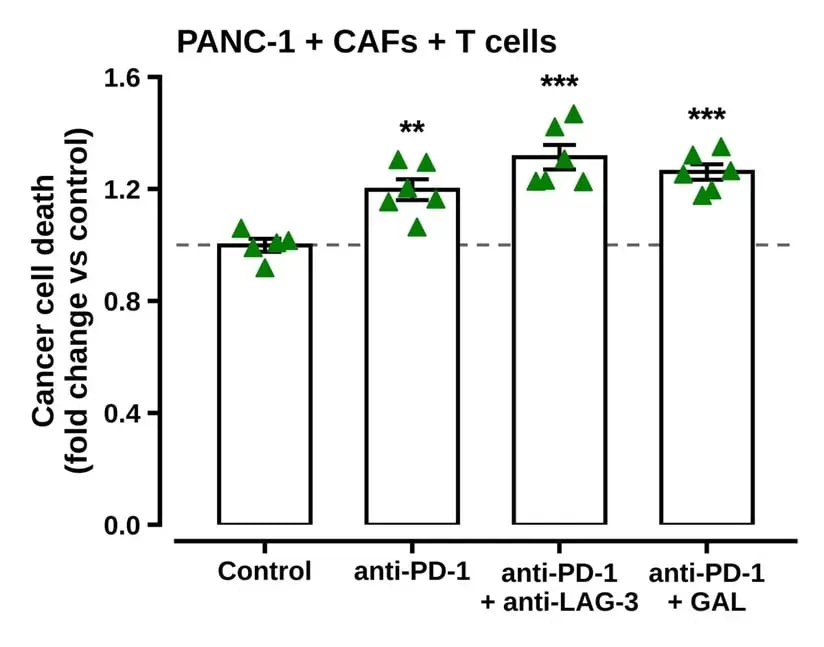
Cancer cell death (fold change versus control) in PDAC-IO tri-cultures 24 hours after treatment with anti-PD-1, anti-PD-1 plus anti-LAG-3, or anti-PD-1 plus galunisertib.
Immunomodulators alter cancer cell death
Twenty-four hours after treatment, tri-cultures exposed to anti-PD-1 alone or in combination with anti-LAG-3 or galunisertib showed higher cleaved caspase-3 signal in pan-cytokeratin-positive cancer cells than cultures treated with an IgG4 isotype control.
These results provide a functional reference for assessing treatment-associated changes in defined tumor-stroma-immune models.
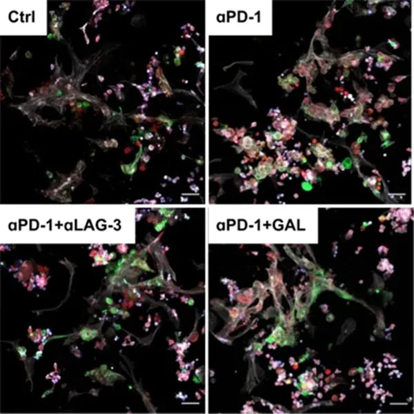
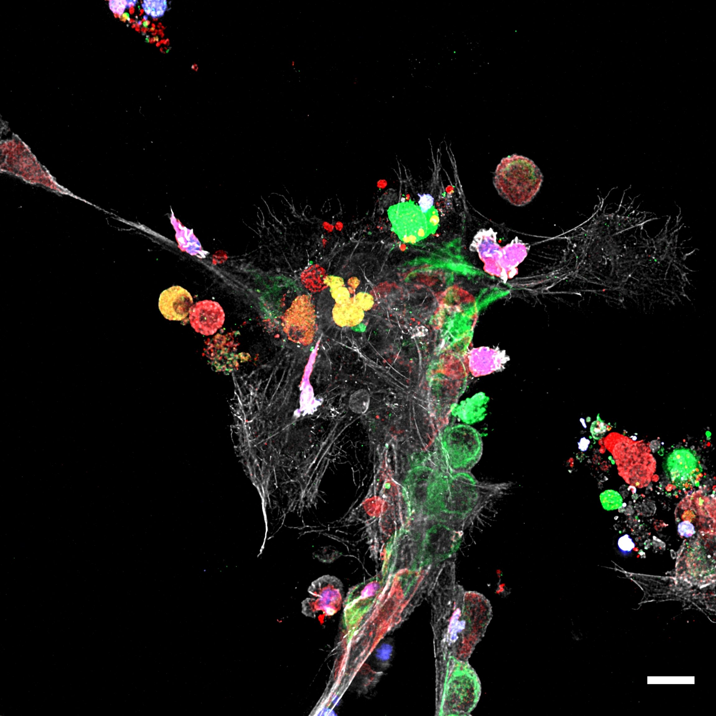
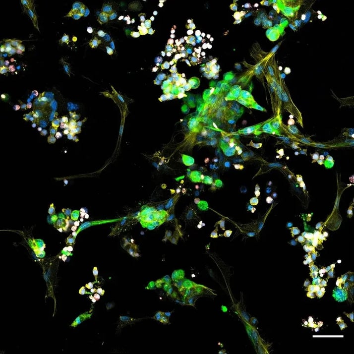
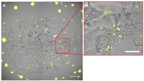
Representative images of PDAC-IO tri-cultures 24 hours after treatment. Pan-cytokeratin (green), cleaved caspase-3 (red), CD8+ T cells (blue) and F-actin (white). Scale bar, 20 µm.
Cancer cell death is measured by imaging
Tri-cultures were treated on day 4 after printing, at the time of CD8+ T cell addition, with anti-PD-1 (10 µg/mL), anti-PD-1 plus anti-LAG-3 (10 µg/mL each), anti-PD-1 plus galunisertib (0.4 µM), or a human IgG4 isotype control. After 24 hours, cultures were fixed and stained for cleaved caspase-3 and pan-cytokeratin to quantify cancer cell death.
What this model demonstrates
The PDAC-IO Validated Solution is designed to support research into tumor-stroma-immune interactions in a defined PDAC context.
-
Tumor, stroma and immune cells are visible together
Cancer cells, CAFs and CD8+ T cells form associations in the hydrogel, and T cells are seen interacting with cancer cells within 24 hours of addition. -
Stroma-associated pathways are enriched
Adding CAFs to PANC-1 and CD8+ T cell cultures is associated with enrichment of epithelial-mesenchymal transition, extracellular matrix and interferon response gene sets. -
Immunomodulators alter cancer cell death
Anti-PD-1, alone or combined with anti-LAG-3 or galunisertib, is associated with higher cancer cell death than an IgG4 isotype control after 24 hours.
Defined model parameters provide a consistent starting point for execution and downstream analysis
-
- Model format: Imaging Model
- Matrix: Px03.84 (about 3 kPa, RGD, YIGSR, GFOGER, HA, full-length fibronectin)
- Cell density: PANC-1 2M/mL; CAFs 10M/mL
- Cell ratio: 1:5 cancer cells:CAFs
- Immune component: 15,000 pre-activated CD8+ T cells per well, added on day 4 after printing
- Culture medium: DMEM : FGM-2 : ImmunoCult (1:1:1) with IL-2 and CD3/CD28 activator
- Treatment timing: Day 4 after printing, with T cell addition, 24 hour exposure
- Reference compounds: Pembrolizumab (anti-PD-1), relatlimab (anti-LAG-3), galunisertib, human IgG4 isotype control
- Downstream analyses: Cleaved caspase-3 and pan-cytokeratin immunofluorescence, optional CellTrace Yellow T cell labeling
Get the PDAC-IO model package
Download the Data Pack
Review supporting data for model setup, tumor-stroma-immune interactions, RNA-seq pathway analysis and checkpoint inhibitor response.
Get the Protocol Pack
Includes protocols for model generation, CD8+ T cell activation, and T cell and treatment addition, plus materials lists, cell conditions, plate plans and expected results.
Need a different context?
Need a different cell source, matrix condition or readout? Talk with Inventia scientists about a custom model.
FAQs
What cells are used, and how are the CD8+ T cells prepared?
The workflow uses PANC-1 pancreatic cancer cells, primary PDAC CAFs and human CD8+ T cells. The T cells are revived on day 1 after printing, activated for three days with a CD3/CD28 activator and IL-2, and added to the models on day 4. Exact cell densities, matrix and treatment conditions are defined in the Protocol Pack.
Is this an antigen-specific T cell killing model?
No. The CD8+ T cells are activated with CD3/CD28 and IL-2 rather than with tumor antigen, so the model is a defined activation context. The functional readout is cancer cell death in the tri-culture, and the workflow does not include a T cell-specific functional readout.
What supporting data are included in the Data Pack?
The Data Pack includes the model specifications, the experimental timeline, representative immunofluorescence images of the tri-culture, bulk RNA-seq pathway analysis comparing tri-cultures with co-cultures of PANC-1 and CD8+ T cells, and the cancer cell-death response to reference treatments. Compartment-resolved RNA-seq deconvolution data are available on request.
What parameters and methods are included in the Protocol Pack?
The Protocol Pack contains three protocols (model generation, CD8+ T cell activation, and T cell and treatment addition) and three supplementary protocols (RNA preparation for sequencing, immunofluorescence staining for cleaved caspase-3 and pan-cytokeratin, and optional CellTrace Yellow labeling of T cells). It also lists materials, cell conditions, plate plans and expected results.
How is PDAC-IO different from the PDAC Validated Solution?
The PDAC Validated Solution is a tumor-CAF co-culture, available with PANC-1 or AsPC-1 cancer cells, built for drug response and stromal chemoprotection studies. PDAC-IO builds on the PANC-1 and CAF model by adding activated CD8+ T cells. Both use the same matrix, cell densities and 1:5 cancer cell-to-CAF ratio. PDAC Validated Solution
Can I use my own cells, CAF source, T cell donor or readout?
Yes. The Validated Solution is a starting point, not a fixed recipe. The supporting data describe the configuration shown: PANC-1 cells, primary PDAC CAFs and human CD8+ T cells in Px03.84. The workflow is modular, so switching in your own cells lets you bring biology specific to your question into the system while likely maintaining an overall biological landscape similar to the one shown. Those data would not apply directly to a customized model, and Inventia Discovery Services can help characterize it.
When should a team use this Validated Solution instead of Discovery Services?
Use the Validated Solution when you want a documented starting point for a question like this one. Use Discovery Services when you need expert help adapting the workflow, for example with different cells, donors, matrices, readouts or treatment contexts, or characterizing a customized model. Discovery Services
Explore Workflow Paths
Browse Validated Solutions
Predefined blueprints for reproducible 3D data in a defined context of use
Explore Discovery Mode
Define and refine 3D cell models to match your biology and readouts
Connect with Discovery Services
Expert support to optimize, validate, and integrate 3D models into discovery workflows